

Article added / Artikel hinzugefügt 01.10.2021
Generally Articles and Discussions about Osteosarcoma in Dogs
→ Evaluations of phylogenetic proximity in a group of 67 dogs with
osteosarcoma: a pilot study
Article added / Artikel hinzugefügt 01.10.2021
Generally Articles and Discussions about Osteosarcoma in Dogs
→ Canine Periosteal Osteosarcoma
Images added / Abbildungen hinzugefügt 02.05.2019
Generally Sonography Atlas of Dogs →
Cardiovascular system → Pulmonary vessels
New subcategory added / Neue Unterkategorie hinzugefügt 02.05.2019
Generally Sonography Atlas of Dogs →
Cardiovascular system → Pulmonary vessels
Images added / Abbildungen hinzugefügt 01.05.2019
Generally Sonography Atlas of Dogs →
Cardiovascular system → Heart valvular diseases


Generally Sonography Atlas of Dogs - Abdomen
(Allgemeiner Sonographie-Atlas von Hunden) - (Abdomen)
Pankreas
(Pankreas)

Ultraschallaufnahme einer Pankreatitis beim Hund
http://de.wikipedia.org/wiki/Pankreatitis
„Illa“ von TA Lizenziert unter Bild-frei über Wikipedia - http://de.wikipedia.org/wiki/Datei:Illa.JPG#mediaviewer/File:Illa.JPG

Pancreatitis in a dog
With special thanks to the authors of the book "Small Animal Ultrasonography" , Marc-André d’Anjou and Dominique Penninck


Contrast-enhanced ultrasonography images of the transverse view of the right pancreatic lobe (p) and mucosa of the descending duodenum adjacent to the right pancreas (d) (both outlined by dotted lines) in 2 representative dogs with pancreatitis (dorsal to the left, ventral to the right, medial to the bottom). (A) Image acquired at 69 seconds after start of contrast agent infusion showing the pancreas at peak intensity. The pancreas is swollen with irregular outlines, and is enhanced nonhomogeneously. Apart from the cranial pancreaticoduodenal vein (v) normally seen, fine pancreatic capillaries are also more prominently enhanced (arrows). Clinical signs of vomiting ceased and activity improved after 4 days of hospitalization and medical treatment. Lipase activity returned to reference range within 10 days. (B) Image acquired at 67 seconds after start of contrast agent infusion showing 2 nonenhancing lesions (arrowheads) in the pancreatic parenchyma. This dog recovered from the acute episode of pancreatitis and was discharged after 10 days of hospitalization and medical treatment. However, lipase activity and C-reactive protein did not return to reference ranges until 46 days and 25 days, respectively.
Lim, S.Y., Nakamura, K., Morishita, K., Sasaki, N., Murakami, M., Osuga, T., Yokoyama, N., Ohta, H., Yamasaki, M. and Takiguchi, M. (2015), Quantitative Contrast-enhanced Ultrasonographic
Assessment of Naturally Occurring Pancreatitis in Dogs. Journal of Veterinary Internal Medicine, 29: 71–78. doi: 10.1111/jvim.12470


A: ultrasound image pancreaticoduodenal vein (arrows) and pancreatic duct ( arrow heads ) in GRMD dog 6 months old. B: A: ultrasound image of pancreaticoduodenal vein ( arrows) in GRMD dog 6 months old (arrows).
Angélica Paula Grando; Arani Nanci Bonfim Mariana; Maria Angélica Miglino; Franklin Almeida Sterman; Mayana Zatz; Luciane Maria Kanayama; Matheus Levi Tjara Feitosa; Daniele dos Santos Martins; Adriana Caroprezo Morini; Juliana Passos Alves dos Santos; Leandro Fadel; Flávio Ribeiro Alves; Carlos Eduardo Ambrósio
"Abdominal and pelvic ultrasonography in healthy golden retriever dogs, carriers and affected by gradual muscular dystrophy" http://dx.doi.org/10.159/S0103-84782008005000037


A transverse ultrasound image through the right lateral abdominal wall with the dog in left ventral recumbency. A round, hypoechoic mass (T) is visible medial to the duodenum (D). This primary pancreatic tumor was not identified with CT and SPECT.
Robbena, J. H., Pollak, Y. W.E.A., Kirpensteijn, J., Boroffka, S. A.E.B., van den Ingh, T. S.G.A.M., Teske, E. and Voorhout, G. (2005), "Comparison of Ultrasonography, Computed Tomography, and Single-Photon Emission Computed Tomography for the Detection and Localization of Canine Insulinoma". Journal of Veterinary Internal Medicine, 19: 15–22. doi: 10.1111/j.1939-1676.2005.tb02652.x

Ultrasound image of the pancreas of a dog with a single pancreatic pseudocyst (black arrows) adjacent to stomach (white arrow). The measurement scale is on the left side of each image; the white horizontal ticks are 1 cm apart.
VanEnkevort, B. A., O'Brien, R. T. and Young, K. M. (1999), "Pancreatic Pseudocysts in 4 Dogs and 2 Cats: Ultrasonographic and Clinicopathologic Findings". doi: 10.1111/j.1939-1676.1999.tb02186.x


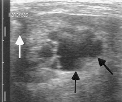
The transverse view of the normal right pancreatic limb (margined by dashed lines) which is located adjacent to the proximal descending duodenum (DUO). Pancreatic duct is indicated by the arrow.
WICKRAMASEKARA RAJAPAKSHAGE, B. K., ELLEARAEWE GARUHAMILAGE, J. P. K., DE SILVA, D. D. N., & DANGOLLA, A. (2016). "Dimensional ultrasonographic relationship of the right lobe of pancreas with associated anatomic landmarks in clinically normal dogs". http://doi.org/10.1292/jvms.15-0209


Ultrasonographic view of the right pancreatic lobe in a clinically healthy dog (arrows indicate organ). Pancreaticoduodenal vein viewed as an anechoic
structure in the pancreas. (d) duodenum. (k) right kidney.
Avante, Michelle L, da Silva, Priscila DA, Feliciano, Marcus AR, Maronezi, Marjury C, Simões, Ana R, Uscategui, Ricardo AR, & Canola, Julio C. (2018). "Ultrasonography of the canine pancreas". Revista MVZ Córdoba, 23(1), 6552-6563. https://dx.doi.org/10.21897/rmvz.1249


Acute pancreatitis in a dog. Enlarged pancreas with reduced echogenicity and increased echogenicity of the adjacent mesentery.
Avante, Michelle L, da Silva, Priscila DA, Feliciano, Marcus AR, Maronezi, Marjury C, Simões, Ana R, Uscategui, Ricardo AR, & Canola, Julio C. (2018). "Ultrasonography of the canine pancreas". Revista MVZ Córdoba, 23(1), 6552-6563. https://dx.doi.org/10.21897/rmvz.1249


Quantitative ARFI ultrasonography of the right pancreatic lobe in a dog.
Avante, Michelle L, da Silva, Priscila DA, Feliciano, Marcus AR, Maronezi, Marjury C, Simões, Ana R, Uscategui, Ricardo AR, & Canola, Julio C. (2018). "Ultrasonography of the canine pancreas". Revista MVZ Córdoba, 23(1), 6552-6563. https://dx.doi.org/10.21897/rmvz.1249
