

Article added / Artikel hinzugefügt 01.10.2021
Generally Articles and Discussions about Osteosarcoma in Dogs
→ Evaluations of phylogenetic proximity in a group of 67 dogs with
osteosarcoma: a pilot study
Article added / Artikel hinzugefügt 01.10.2021
Generally Articles and Discussions about Osteosarcoma in Dogs
→ Canine Periosteal Osteosarcoma
Images added / Abbildungen hinzugefügt 02.05.2019
Generally Sonography Atlas of Dogs →
Cardiovascular system → Pulmonary vessels
New subcategory added / Neue Unterkategorie hinzugefügt 02.05.2019
Generally Sonography Atlas of Dogs →
Cardiovascular system → Pulmonary vessels
Images added / Abbildungen hinzugefügt 01.05.2019
Generally Sonography Atlas of Dogs →
Cardiovascular system → Heart valvular diseases


Generally Sonography Atlas of Dogs - Abdomen
(Allgemeiner Sonographie-Atlas von Hunden) - (Abdomen)
Gallbladder
(Gallenblase)

Canine normal liver, gallbladder, spleen
(from Nottingham Vet School)


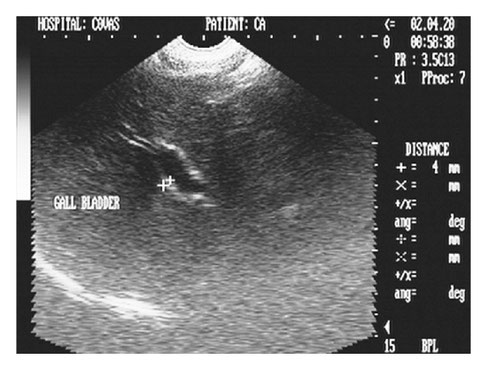
Gall-Bladder in a Dachshund-Mix
Thomas Echter / Vanessa Ermer


A: image of ultra-sonografphy of the gall bladder with presenca of calculations in his interior in dog GRMD of 6 months of age.
B: image of ultra-sonography of the gall bladder with presenca of dense bile in dog adult GRMD.
Angélica Paula Grando; Arani Nanci Bonfim Mariana; Maria Angélica Miglino; Franklin Almeida Sterman; Mayana Zatz; Luciane Maria Kanayama; Matheus Levi Tjara Feitosa; Daniele dos Santos Martins; Adriana Caroprezo Morini; Juliana Passos Alves dos Santos; Leandro Fadel; Flávio Ribeiro Alves; Carlos Eduardo Ambrósio
"Abdominal and pelvic ultrasonography in healthy golden retriever dogs, carriers and affected by gradual muscular dystrophy" http://dx.doi.org/10.159/S0103-84782008005000037


Irregular wall thickening of the gall- bladder with increased echogenicity.
Corina F Guendulain, MV; Griselda M González, MV, Esp Cs Cl Vet; Carmen I Maffrand, MV, Esp Cs Cl Vet.
"The ultrasonography as an assistance to diagnostic of cholecystitis in a canine"
Rev Colom Cienc Pecua vol.23 no.1 Medellín Jan./Mar. 2010


Transverse cutting of the gallbladder . Hyperechoic aggregation of bile in the bottom of the gallbladder is displayed .
Corina F Guendulain, MV; Griselda M González, MV, Esp Cs Cl Vet; Carmen I Maffrand, MV, Esp Cs Cl Vet.
"The ultrasonography as an assistance to diagnostic of cholecystitis in a canine"
Rev Colom Cienc Pecua vol.23 no.1 Medellín Jan./Mar. 2010


Longitudinal view of the gallbladder . Arrowheads point to the external and internal echogenic serous mucous edges.
Corina F Guendulain, MV; Griselda M González, MV, Esp Cs Cl Vet; Carmen I Maffrand, MV, Esp Cs Cl Vet.
"The ultrasonography as an assistance to diagnostic of cholecystitis in a canine"
Rev Colom Cienc Pecua vol.23 no.1 Medellín Jan./Mar. 2010


Gallbladder inflammation and liver (cholangitis and cholangiohepatitis):
Images of two dogs with hepato-megaly, increased hepatic echo- genicity, cystic duct dilation and gallbladder thickening , showing a cholangiohepatitis.
With special thanks to Priscilla Pinel, Medical Veterinary.
Currently serves in veterinary clinics and homes for the municipality of Rio de Janeiro ( south, north and west ).
http://veterinariapriscillapinel.com.br


Gallbladder inflammation and liver (cholangitis and cholangiohepatitis): Images of two dogs with dilated cystic duct , the second still with thickening of the gallbladder wall , showing a cholangitis.
With special thanks to Priscilla Pinel, Medical Veterinary.
Currently serves in veterinary clinics and homes for the municipality of Rio de Janeiro ( south, north and west ).
http://veterinariapriscillapinel.com.br


Chronic cholangiohepatitis.
With special thanks to Priscilla Pinel, Medical Veterinary.
Currently serves in veterinary clinics and homes for the municipality of Rio de Janeiro ( south, north and west ).
http://veterinariapriscillapinel.com.br


Bile mud ( sediment accumulation in the bladder).
With special thanks to Priscilla Pinel, Medical Veterinary.
Currently serves in veterinary clinics and homes for the municipality of Rio de Janeiro ( south, north and west ).
http://veterinariapriscillapinel.com.br


Ultrasonography of gallbladder stones.
The stones in the gallbladder were a finding in routine ultrasound study of a bitch (Foxterrier race) , 13 years old.
With special thanks to Irene García Patiño (Sombra Acústica), veterinarian at the Veterinary Clinic Argos in Cee (A Coruña, Spain). http://sombraacustica.com


Cross-sectional image of mucocele dog gallbladder with a starry pattern and bile anechoic around.
Fred S. Pike, John Berg, Norval W. King, Dominique G. Penninck, Cynthia R. L. Webster
"MUCOCELE GALLBLADDER in dogs: 30 cases (2000-2002)"


Slitting of a ruptured mucocele dog gallbladder. F: free peritoneal fluid. St: hyperechoic abdominal fat.
Fred S. Pike, John Berg, Norval W. King, Dominique G. Penninck, Cynthia R. L. Webster
"MUCOCELE GALLBLADDER in dogs: 30 cases (2000-2002)"


Ultrasonographic image (8 MHz convex array probe, Esaote, MyLab, Genoa, Italy) of a sagittal section of a gallbladder in a dog diagnosed with a gallbladder mucocoele.
Smalle, T.M., Cahalane, A.K. & Köster, L.S., 2015, ‘Gallbladder mucocoele: A review’, http://dx.doi.org/10.4102/jsava.v86i1.1318


Ultrasonographic image (8 MHz convex array probe, Esaote, MyLab, Genoa, Italy) of a sagittal section of a gallbladder in a dog diagnosed with a gallbladder mucocoele.
Smalle, T.M., Cahalane, A.K. & Köster, L.S., 2015, ‘Gallbladder mucocoele: A review’, http://dx.doi.org/10.4102/jsava.v86i1.1318


Sonograph (2D) in transverse scan showing hyperechoic liver lobe and cholecystitis with inspissated bile in gall bladder (GB) in a 7-year-old intact Pomeranian male dog.
Kumar V, Kumar A, Varshney AC, Tyagi SP, Kanwar MS, Sharma SK. Diagnostic Imaging of Canine Hepatobiliary Affections: A Review. Veterinary Medicine International. 2012;2012:672107. doi:10.1155/2012/672107.


Transverse sonogram (2D) of right medial liver lobe of two and half-year -old intact female Pointer dog depicting generalized hyperechoic parenchyma with biliary sludge in gall bladder.
Kumar V, Kumar A, Varshney AC, Tyagi SP, Kanwar MS, Sharma SK. Diagnostic Imaging of Canine Hepatobiliary Affections: A Review. Veterinary Medicine International. 2012;2012:672107. doi:10.1155/2012/672107.


Hepatic sonogram (2D) in sagittal scan of a five-year-old intact Pomeranian male dog depicting increased paren-chymal echogenicity with hyperechoic double rim appearance of GB wall.
Kumar V, Kumar A, Varshney AC, Tyagi SP, Kanwar MS, Sharma SK. Diagnostic Imaging of Canine Hepatobiliary Affections: A Review. Veterinary Medicine International. 2012;2012:672107. doi:10.1155/2012/672107.

Two-dimensional ultrasonographic appearance in sagittal scan of cirrhosed liver lobe in a seven-year-old intact mixed-breed dog depicting collapsed and irregularly shaped gall bladder with echogenic and thickened (4 mm) GB wall.
Kumar V, Kumar A, Varshney AC, Tyagi SP, Kanwar MS, Sharma SK. Diagnostic Imaging of Canine Hepatobiliary Affections: A Review. Veterinary Medicine International. 2012;2012:672107. doi:10.1155/2012/672107.


Longitudinal sonogram (2D) of liver of two and half-year-old, intact female Pointer dog showing cholecystitis as echogenic thickened GB wall and mineralization in the neck of gall bladder with acoustic shadowing along with hyperechoic parenchyma.
Kumar V, Kumar A, Varshney AC, Tyagi SP, Kanwar MS, Sharma SK. Diagnostic Imaging of Canine Hepatobiliary Affections: A Review. Veterinary Medicine International. 2012;2012:672107. doi:10.1155/2012/672107.
