Article added / Artikel hinzugefügt 01.10.2021
Generally Articles and Discussions about Osteosarcoma in Dogs
→ Evaluations of phylogenetic proximity in a group of 67 dogs with
osteosarcoma: a pilot study
Article added / Artikel hinzugefügt 01.10.2021
Generally Articles and Discussions about Osteosarcoma in Dogs
→ Canine Periosteal Osteosarcoma
Images added / Abbildungen hinzugefügt 02.05.2019
Generally Sonography Atlas of Dogs →
Cardiovascular system → Pulmonary vessels
New subcategory added / Neue Unterkategorie hinzugefügt 02.05.2019
Generally Sonography Atlas of Dogs →
Cardiovascular system → Pulmonary vessels
Images added / Abbildungen hinzugefügt 01.05.2019
Generally Sonography Atlas of Dogs →
Cardiovascular system → Heart valvular diseases


Generally Sonography Atlas of Dogs - Abdomen
(Allgemeiner Sonographie-Atlas von Hunden) - (Abdomen)
Liver - Page 1
(Leber - Seite 1)


Normal picture of the liver . VB = gallbladder ; PV = portal vein ; VH = hepatic vein ; D = membrane .
N. Díez Bru
"Ecografía abdominal en pequeños animales."
CLINICA VETERINARIA DE PEQUEÑOS ANIMALES
Volumen 12, Número 3, Julio/Septiembre 1992


The liver is hypoechoic foci and
heterogeneous parenchymal echogenicity . A = ascites ; H = hígdo , Histopathological diagnosis : Cirrosis of the liver
N. Díez Bru
"Ecografía abdominal en pequeños animales."
CLINICA VETERINARIA DE PEQUEÑOS ANIMALES
Volumen 12, Número 3, Julio/Septiembre 1992


The liver is hypoechoic foci , A = ascitú ; H = liver , Histopathological diagnosis : undifferentiated sarcoma liver
N. Díez Bru
"Ecografía abdominal en pequeños animales."
CLINICA VETERINARIA DE PEQUEÑOS ANIMALES
Volumen 12, Número 3, Julio/Septiembre 1992

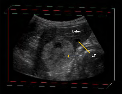
Erfasster Rohdatensatz im Würfelmodell, Leber mit reflexreichem und reflexarmen Lebertumor (LT)
Peppler, Christine "Dreidimensionale Sonographie der Leber beim Hund", 2007
URL: http://geb.uni-giessen.de/geb/volltexte/2007/4704


Bewegungsartefakt im Würfelmodell
Peppler, Christine "Dreidimensionale Sonographie der Leber beim Hund", 2007
URL: http://geb.uni-giessen.de/geb/volltexte/2007/4704


Bewegung der coronaren Ebene, Leberzyste

Bewegung der sagittalen Ebene, Leberzyste
Bewegung der sagittalen Ebene, gewebige Zubildung (LT: Lebertumor)
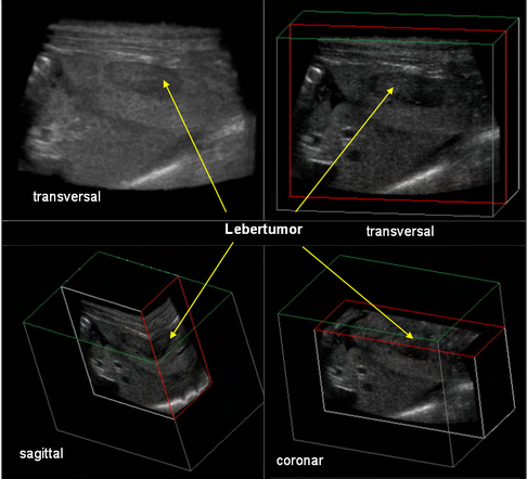
Gleichzeitige Würfeldarstellung mit Funktion Tile, gleichzeitige Darstellung von B-Bild und der verschobenen transversalen, sagittalen und coronaren Ebene eines Lebertumors

Status Main Darstellung, gleichzeitige Darstellung einer Ebene im coronaren, sagittalen und transversalen Schnitt einer Leberzyste.
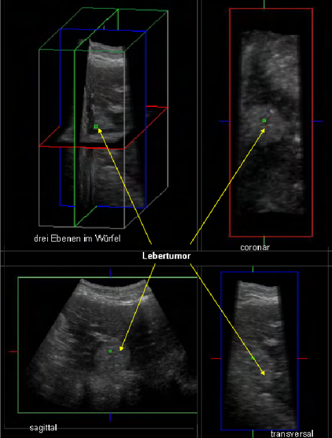
Status Main Darstellung, B-Bild, coronare, sagittale und transversale Ebene eines Lebertumors.

Parallelmodus, gleichzeitige Darstellung einer Leberzyste mit vier Schnittbildern.
Peppler, Christine "Dreidimensionale Sonographie der Leber beim Hund", 2007
URL: http://geb.uni-giessen.de/geb/volltexte/2007/4704


Typ 1 Grey Surface, keine Einstellung Typ 2, Darstellung eines Lebertumors.

Typ 1 Grey Surface, Typ 2 Texture, Darstellung eines Lebertumors (LT).
Peppler, Christine "Dreidimensionale Sonographie der Leber beim Hund", 2007
URL: http://geb.uni-giessen.de/geb/volltexte/2007/4704


Artefakt durch Gasansammlung im Darm, LT (Lebertumor)

Typ 1 Texture, Typ 2 Grey Surface mit 2D Artefakt, Darstellung eines Lebertumors (LT) mit Überlagerung von Darm mit Gasartefakt.

Typ 1 Grey Surface, Typ 2 Texture mit 2D Artefakt, Darstellung eines Lebertumors (LT) mit Überlagerung von Darm mit Gasartefakt.
Peppler, Christine "Dreidimensionale Sonographie der Leber beim Hund", 2007
URL: http://geb.uni-giessen.de/geb/volltexte/2007/4704


Typ 1 Texture, Typ 2 Grey Surface, grosser reflexreicher und kleiner reflexarmer Lebertumor (LT).

Typ 2 Texture, Typ 1 Grey Surface, grosser reflexreicher und kleiner reflexarmer Lebertumor (LT).

Typ 1 Grey Surface, keine Einstellung Typ 2, grosser reflexreicher und kleiner reflexarmer Lebertumor (LT).
Peppler, Christine "Dreidimensionale Sonographie der Leber beim Hund", 2007
URL: http://geb.uni-giessen.de/geb/volltexte/2007/4704


Einstellung der Tile-Funktion, gleichzeitige Darstellung eines gerenderten Bildes und eines Würfelmodells, Lebertumor (LT), Leberzyste (LZ).

Volumetrie mit Serial-Methode.
Peppler, Christine "Dreidimensionale Sonographie der Leber beim Hund", 2007
URL: http://geb.uni-giessen.de/geb/volltexte/2007/4704


Kategorie 1 der Parenchymveränderung (Lebertumor), Gallenblase (GB) mit abgebildet.

Kategorie 2 der Parenchymveränderung (Leberzyste LZ), Gallenblase (GB) mit abgebildet.

Kategorie 3 der Parenchymveränderung (LZ: Leberzyste).
Peppler, Christine "Dreidimensionale Sonographie der Leber beim Hund", 2007
URL: http://geb.uni-giessen.de/geb/volltexte/2007/4704


Zwerchfellnahe, rechtsseitige Leberzyste (LZ), Gallenblase (GB).
Peppler, Christine "Dreidimensionale Sonographie der Leber beim Hund", 2007
URL: http://geb.uni-giessen.de/geb/volltexte/2007/4704


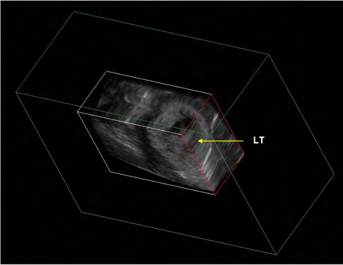
Randständig, rechtsseitiger Lebertumor (LT), Gallenblase (GB).
Peppler, Christine "Dreidimensionale Sonographie der Leber beim Hund", 2007
URL: http://geb.uni-giessen.de/geb/volltexte/2007/4704


Zentraler, linksseitiger Lebertumor (LT).
Peppler, Christine "Dreidimensionale Sonographie der Leber beim Hund", 2007
URL: http://geb.uni-giessen.de/geb/volltexte/2007/4704


Canine normal liver, gallbladder, spleen
(from Nottingham Vet School)


Ultrasonographic assessment of gastric motility. Cross-sectional view of the gastric antrum (arrows) at 1 min (A) and 15 min (B) after ingestion of the consommé soup.
Daisuke Kamino, Noriaki Manabe, Jiro Hata, Ken Haruma, Shinji Tanaka, Kazuaki Chayama
1 March 2008
2266051
Journal of Clinical Biochemistry and Nutrition
the Society for Free Radical Research Japan


Initial hepatic ultrasonographic image. An oblique plane is used with a ventral acoustic window, showing part of the gallbladder (white star), rounded liver margin (black arrow) and heterogenous parenchyma.
Two – days post-biopsy hepatic ultarsonographic image. Approximately the same plane is used and the same acoustic window as Figure 1, revealing echoic gas foci (black arrowheads) at the previous biopsy site. Gallbladder (white star) shows a normal appearance.
"Hepatic emphysema associated with ultrasound-guided liver biopsy in a dog"
Frida Westgren, Tove Hjorth, Margareta Uhlhorn, Pernille E Etterlin and Charles J Ley
doi:10.1186/1751-0147-56-25
http://www.actavetscand.com


Graphische Darstellung der Ankunft des Kontrastmittels. Links im Bild die farblich unterschiedlichen ROI`s (gelb auf der Arterie und grün über einer Lebervene) im B-Bild. Auf der rechten Seite die Graphik mit dem Grad der Verstärkung in dB (y-Achse) über die Zeit in Sekunden (x-Achse). Es zeigt sich ein steiler Anstieg der Verstärkung nach 10s in der Arterie (gelb), ein kurzer Peak und anschließend schnelles Abfluten. In der Lebervene zeigt sich nach ca.20s ein langsamer Anstieg der Verstärkung durch die Ankunft des Kontrastmittels
Antje Trogisch-Hause, "Die Hepatische Transitzeit des
Echosignalverstärkers SonoVue® beim Hund", Veterinärmedizinische Fakultät Universität Leipzig
http://www.qucosa.de/fileadmi/data/qucosa/documen/ 7838/DruckversionEnd_Trogisch-Hause13.10.2011.pdf


Duplexbild (B-Modus und PW-Doppler) einer Lebervene. Im oberen B-Bild zeigt sich die interkostale Anschallung der Leber mit Lebervenenästen. Der Spektraldoppler zeigt das typische triphasische Lebervenenmuster. Der erste Geschwindigkeitsgipfel ist zur V.cava hin gerichtet entsteht durch dieVorhof- systole. Die zweite Welle zeigt die atriale Füllung- sphase. Das Blut entleert sich aus der Lebervene in die V.cava. Die dritte Welle ist während dem Blutabfluss aus dem Vorhof in den rechten Ventrikel. In der nachfolgenden Füllungsphase steigt der atriale Durck und die Geschwindigkeit nimmt wieder ab.
Antje Trogisch-Hause, "Die Hepatische Transitzeit des
Echosignalverstärkers SonoVue® beim Hund", Veterinärmedizinische Fakultät Universität Leipzig
http://www.qucosa.de/fileadmi/data/qucosa/documen/ 7838/DruckversionEnd_Trogisch-Hause13.10.2011.pdf


TIC-Analyse der Ankunftzeiten des Kontrastmittels in einer Leberarterie und einer Lebervene. Die
Abbildung zeigt objektive Erfassung der Ankunftszeiten in den
Lebergefäßen. Links im Bild befinden sich die 1mm g
roßen ROI's über den Gefäßen. Im Diagramm rechts zeigt sich der Kontrastmittelverlauf in der Leberarterie (gelb). Bei Ankunft des
Kontrastmittels zeigt sich ein steiler Anstieg der Verstärkung, ein spitzer Peak und
ein schnelles wieder abfluten. Im Gegensatz dazu zeigt sich das zeitlich spätere Anfluten des Kontrastmittels in einer Lebervenen (grün). Die Kurve der
Verstärkung durch das Kontrastmittel verläuft flacher.
Antje Trogisch-Hause, "Die Hepatische Transitzeit des
Echosignalverstärkers SonoVue® beim Hund", Veterinärmedizinische Fakultät Universität Leipzig
http://www.qucosa.de/fileadmi/data/qucosa/documen/ 7838/DruckversionEnd_Trogisch-Hause13.10.2011.pdf


Parenchymas difference in two animals ; the first image of a hyperechoic liver ( diffuse echogenicity ) with severe biliary sludge in the gallbladder , the following two images are of a hypoechoic liver of a dog suspected of hemoparasitosis and Alkaline Phosphatase outside the box limits
With special thanks to Priscilla Pinel, Medical Veterinary.
Currently serves in veterinary clinics and homes for the municipality of Rio de Janeiro ( south, north and west ).
http://veterinariapriscillapinel.com.br


Rounded hypoechoic liver nodules , located in the left lobe of a canine 9 years.
With special thanks to Priscilla Pinel, Medical Veterinary.
Currently serves in veterinary clinics and homes for the municipality of Rio de Janeiro ( south, north and west ).
http://veterinariapriscillapinel.com.br


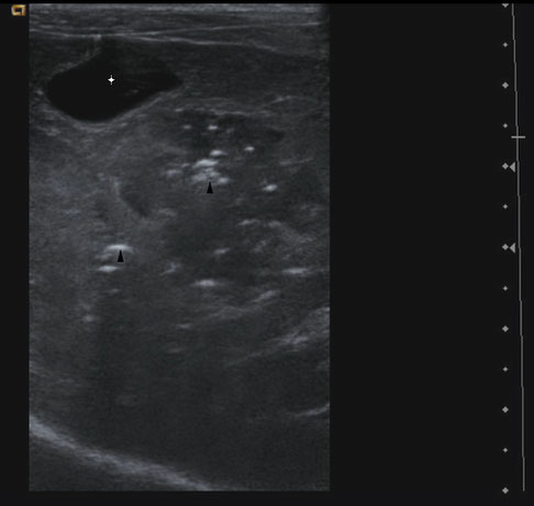
Canine chronic hepatitis.
Canine 6 years with increased echogenicity and diffuse hyperechoic nodules , and her blood test found an increase of ALT and Phosphatase.
With special thanks to Priscilla Pinel, Medical Veterinary.
Currently serves in veterinary clinics and homes for the municipality of Rio de Janeiro ( south, north and west ).
http://veterinariapriscillapinel.com.br