Article added / Artikel hinzugefügt 30.03.2018
"Prognostic factors of early metastasis and mortality in dogs with appendicular osteosarcoma after receiving surgery: An individual patient data meta-analysis"
Article added / Artikel hinzugefügt 26.03.2018
"Identification of myeloid derived suppressor cells in the peripheral blood of tumor bearing dogs"
Article added / Artikel hinzugefügt 10.12.2017
"Clinical and Immunomodulatory Effects of Toceranib Combined with Low-Dose Cyclophosphamide in Dogs with Cancer"
Article added / Artikel hinzugefügt 10.12.2017
"A Comparison of Five Different Bone Resorption Markers in Osteosarcoma-Bearing Dogs, Normal Dogs, and Dogs with Orthopedic Diseases"
Article added / Artikel hinzugefügt 10.12.2017
"Decreased Ratio of CD8+ T Cells to Regulatory T Cells Associated with Decreased Survival in Dogs with Osteosarcoma"
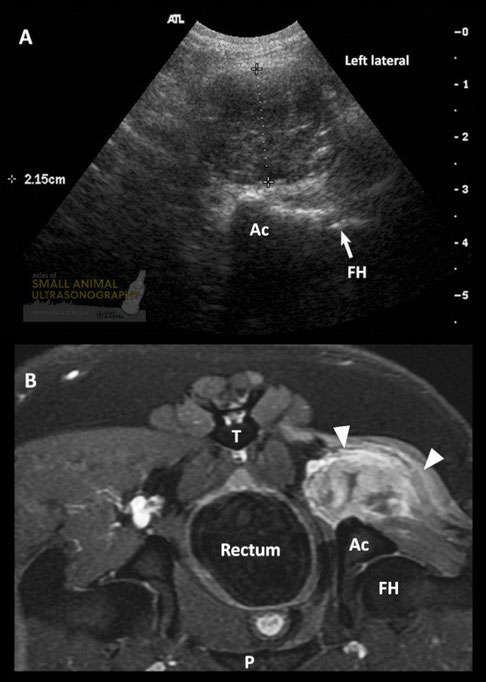
Sciatic nerve sheath fibrosarcoma in a dog. A: Transverse sonographic image of a 2.15cm thick hypoechoic mass located dorsal to the acetabular bone (Ac) and femoral head (FH). B: Transverse post-contrast T1-weighted MR sequence with fat saturation, highlighting the contrast-enhancing mass (arrowheads). The adjacent muscles are atrophied and hyperintense. T, tail; P, pubis.
With special thanks to the authors of the book "Small Animal Ultrasonography" , Marc-André d’Anjou and Dominique Penninck
Primary flexor enthesopathy. Medial (A) and dorsal (B) views of the elbow of a 14 month-old Golden retriever dog produced with computed tomographic (CT) volume-rendering, with the overlying transverse and longitudinal planes used to produce the sonographc images on the right (respectively). Large (M) and small (m) mineral fragments are depicted in all images. These mineral bodies are embedded in a heterogeneous mass of moderate echogenicity (arrowheads) consistent with fibrous and metaplastic soft tissue, in part confluent with some of the flexor tendons (T). Note the stronger acoustic enhancement associated with the larger mineral bodies.
With special thanks to the authors of the book "Small Animal Ultrasonography" , Marc-André d’Anjou and Dominique Penninck
Fragmentation of the medial coronoid process. A: Medial view of the elbow of a 12 month-old Labrador retriever dog produced with computed tomographic (CT) volume-rendering, on which the fragmentation of the medial coronoid process (FMCP) is well visualized. B: This longitudinal sonographic image was obtained with the probe placed medially on the joint, the marker oriented proximally. The joint capsule is distended and thickened (arrowhead) and a gap (arrow) is present at the distal aspect of the medial coronoid process (MCP). The humeral condyle (HC) is mildly irregular. C and D: Corresponding transverse CT (C) and sonographic (D) images in which a fissure (arrows) extends through the MCP. The joint capsule is distended (arrowhead). R, radial head. U, ulna.
With special thanks to the authors of the book "Small Animal Ultrasonography" , Marc-André d’Anjou and Dominique Penninck
Joint tumor in a dog
With special thanks to the authors of the book "Small Animal Ultrasonography" , Marc-André d’Anjou and Dominique Penninck
Contrast enhanced ultrasonography on harmonic detection mode before (A) and after (B) contrast agent injection at the skeletal muscle of the humerus. Transient increase of vascular intensity (arrows) and then disappearance was easily observed from all skeletal muscles after injecting the contrast medium.
Juyeon OH, Sunghoon JEON, and Jihye CHOI
Conventional ultrasonography on color Doppler mode (A) and contrast enhanced ultrasonography on harmonic detection mode (B) of radius. Region of interest placed over the muscle on contrast enhanced ultrasonography and time‒intensity curves (C). The muscle area had a typical parabolic curve, which consisted of inflow, outflow and peak intensity.
Juyeon OH, Sunghoon JEON, and Jihye CHOI
Imagen ecográfica correspondient e al corte sagital de la región suprarrotuliana en la que se visualiza el tendón del músculo cuádriceps femoral con estructura fibrilar y peritendón hiperecogénico (flechas). P: proximal, D: distal, TF: tróclea del fémur.
Ultrasound image correspondient and the sagittal section of the region suprapatellar in which the tendon of the quadriceps femoris muscle displayed c on peritendon hyperechoic fibrillar structure ( arrows). Q: proximal , D : distal , TF : trochlea of the femur.
Soler Laguía, Marta
"Diagnóstico por imagen de la articulación de la rodilla en la especie canina"
oai:digitum.um.es:10201/40906
Imagen ecográfica del corte sagital de la regi
ón suprarrotuliana en la que se visualiza la superficie
convexa hiperecogénica de la rótula (flechas). P: proximal, D: distal, R: rótula.
Sagittal ultrasound image of the region suprapatellar ng
displayed in the hyperechoic convex surface of the patella ( arrows). Q: proximal , D : distal , R :
kneecap.
Soler Laguía, Marta
"Diagnóstico por imagen de la articulación de la rodilla en la especie canina"
oai:digitum.um.es:10201/40906
Imagen ecográfica del corte sagital de la región
suprarrotuliana en la que visualizamos el cartílago articular de la tróclea del fémur, como una línea hipoecogénica lisa
demarcada por dos interfases
hiperecogénicas (flechas). P: proximal, D: distal, R: rótula, F: fémur.
Sagittal ultrasound image of the resuprapatellar region in
which we visualize the articular cartilage of the trochlea
l femur, as a hypoechoic smooth line demarcated by two interfaces hyperechoic ( arrows). Q: proximal , D : distal , R : patella, F: femur.
Soler Laguía, Marta
"Diagnóstico por imagen de la articulación de la rodilla en la especie canina"
oai:digitum.um.es:10201/40906
Imagen ecográfica correspondiente al corte sagital del ligamento rotuliano, que se visualiza con estructura fibrilar y peritendón
hiperecogénico (flechas). P: proximal, D: distal, R: rótula, TF: tróclea del fémur.
Ultrasound image corresponding to the sagittal section of the patellar ligament, which is displayed with hyperechoic fibrillar
structure and peritendon ( arrows). Q: proximal , D: distal, R : patella TF: trochlea of the
femur.
Soler Laguía, Marta
"Diagnóstico por imagen de la articulación de la rodilla en la especie canina"
oai:digitum.um.es:10201/40906
Imagen ecográfica del corte sagital de la región infrarrotuliana en la que se observa el cuerpo adiposo infrarrotuliano, caudal al
ligamento rotuliano con una ecogenicidad media homogénea (flechas). P: proximal, D: distal, F: fémur, T: tibia, LR: ligamento rotuliano.
Sagittal ultrasound image of the infrapatellar region where the infrapatellar fat pad is observed flow to the patellar ligament
with a homogeneous medium echogenicity ( arrows). Q: proximal , D : distal , F: femur, T: warm , LR : patellar ligament.
Soler Laguía, Marta
"Diagnóstico por imagen de la articulación de la rodilla en la especie canina"
oai:digitum.um.es:10201/40906
Imagen ecográfica de un corte longitudinal sagital de la región infrarrotuliana en la que se visualiza el ligamento cruzado
craneal (flechas) extendiéndose desde el área intercondilar central de la tibia hasta la fosa intercondilar del fémur. P: proximal, D: distal, F: fémur, T: tibia.
Ultrasound image of a sagittal Slitting infrapatellar region where the cranial cruciate ligament ( arrows ) extending from the
central intercondylar area of the tibia to the intercondylar fossa of the femur is displayed . Q:proximal , D : distal , F:
femur, T: tibia.
Soler Laguía, Marta
"Diagnóstico por imagen de la articulación de la rodilla en la especie canina"
oai:digitum.um.es:10201/40906
Imagen ecográfica correspondiente a un corte sagital de la
región infrarrotuliana en la que se pueden observar los dos ligamentos cruzados craneal (flecha azul) y caudal
(fl
echa amarilla) formando una “V” y presentando la misma ecogenicidad. P: proximal, D: distal, F: fémur, T: tibia.
Corresponding ultrasound image a sagittal section of the
region infrapatellar where you can see the two cross head (blue arrow ) ligaments and flow (fl yellow arrow) forming a " V " and presenting the same echogenicity. P :
proximal , D : distal , F: femur, T: tibia.
Soler Laguía, Marta
"Diagnóstico por imagen de la articulación de la rodilla en la especie canina"
oai:digitum.um.es:10201/40906
Imagen ecográfica de un corte sagital de la región infrarrotuliana en la que se visualiza el ligamento cruzado caudal, desde el
punto de fijación de la superficie lateral del cóndilo femoral medial, dirigiéndose paralelo a éste, hacia la escotadura poplítea de la tibia (flecha). P: proximal, D: distal, F: fémur, T: tibia.
Ultrasound image of a sagittal section of the infrapatellar region where the cruciate ligament is displayed flow , from the point
of attachment of the side surface of the medial femoral condyle , addressing parallel thereto , to the pop recess Litea
tibia ( arrow ) . Q: proximal , D : distal , F: femur, T: tibia.
Soler Laguía, Marta
"Diagnóstico por imagen de la articulación de la rodilla en la especie canina"
oai:digitum.um.es:10201/40906
Corte sagital de la región infrarrotuliana en la que se observa el cartílago articular del cóndilo lateral del fémur
hipoecogénico, demarcado por dos líneas hiperecogénicas lisas, paralelas a la superficie del cóndilo (flechas). P: proximal, D: distal, F: fémur.
Sagittal section of the infrapatellar region in which articular cartilage is observed lateral condyle of hypoechoic femur ,
demarcated by two parallel to the surface of the condyle (arrows) hyperechoic smooth lines . Q: proximal , D : distal , F: femur.
Soler Laguía, Marta
"Diagnóstico por imagen de la articulación de la rodilla en la especie canina"
oai:digitum.um.es:10201/40906
Imagen ecográfica del corte sagital de la región lateral en la que se visualiza el tendón de origen del músculo extensor digital
largo delimitado por el peritendón hiperecogénico (flechas azules), localizándose inmediatamente
lateral y superficial al menisco lateral (flechas rojas). P: proximal, D: distal, F: fémur, T:
tibia.
Sagittal ultrasound image of the lateral region in which the tendon origin of extensor digitorum longus muscle delimited by the
hyperechogenic peritendon (blue arrows) is displayed ,being located immediately latera
lateral meniscus surface to ly ( red arrows) . Q: proximal , D : distal , F: femur, T: tibia.
Soler Laguía, Marta
"Diagnóstico por imagen de la articulación de la rodilla en la especie canina"
oai:digitum.um.es:10201/40906
Imagen ecográfica correspondiente al corte sagital de la región medial en la que observamos el menisco medial como una estructura
hipoecogénica homogénea con forma triangular (flechas). P: proximal, D: distal, F: fémur, T:
tibia.
Ultrasound image corresponding to the sagittal section of the medial region in which we observe the medial meniscus as a
homogeneous hypoechoic triangular structure ( arrows). P :proximal , D : distal , F: femur, T:
tibia.
Soler Laguía, Marta
"Diagnóstico por imagen de la articulación de la rodilla en la especie canina"
oai:digitum.um.es:10201/40906
Ecografia transversa del surco bicipital de un perro con artrosis. Se visualiza un osteofito (flecha)
incidiendo en el tendón del bíceps e irregularidades en el surco (puntas de.flecha).
Transverse ultrasound biceps groove of a dog with osteoarthritis. An osteophyte (arrow ) focusing on the biceps tendon and
irregularities in the groove ( de.flecha tips) is displayed .
I. Gielen
"Comparison of Imaging Techniques used in the Osteoarthritic Joint and their Interpretation"
Clin. Vet. Peq. Anim, 26 (2): 137-144,2006
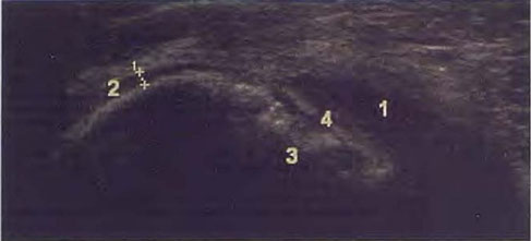
Ecografia de un colgajo de
OCD den la zona de la cabeza humeral caudal de un perro. 1=líquido articular; 2=cartilago
articular sano; 3=cráter subcondral irregular; 4=línea hiperecoica patognomónica de la presencia de un colgajo.
Ultrasound of a flap
OCD den area flow of the humeral head of a dog. 1 = joint fluid ; 2 = healthy articular cartilage ; 3 = subchondral irregular
crater regulate ; 4 = line hyperechoic pathognomonic presence of a flap.
I. Gielen
"Comparison of Imaging Techniques used in the Osteoarthritic Joint and their Interpretation"
Clin. Vet. Peq. Anim, 26 (2): 137-144,2006
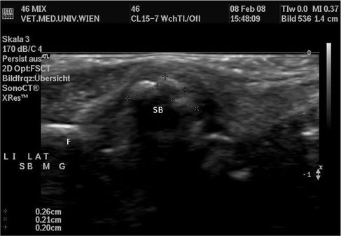
Sesambeinteilung, Havaneser, weibl. 13 Jahre , Sagittalschnitt durch das lat. SB der li. Seite (SB: Sesambeinanteile, F: Femur)
Stefan Perterer
"Sonographie, Röntgenologie und Pathologie des Musculus gastrocnemius im Ursprungs-bereich beim Hund"
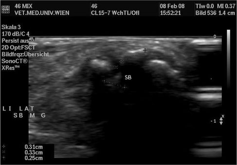
Sesambeinteilung, Havaneser, weibl. 13 Jahre , Transversalschnitt durch das lat. SB der li. Seite (SB: Sesambeinanteile)
Stefan Perterer
"Sonographie, Röntgenologie und Pathologie des Musculus gastrocnemius im Ursprungs-bereich beim Hund"
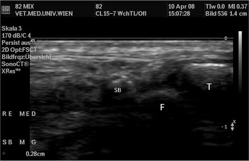
Verlagerung, Mischling, weibl. 8 Jahre , Sagittalschnitt durch das med. SB der re. Seite (SB: Sesambein, F: Femur, T: Tibia)
Stefan Perterer
"Sonographie, Röntgenologie und Pathologie des Musculus gastrocnemius im Ursprungs-bereich beim Hund"
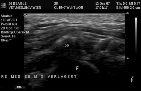
Verlagerung, Beagle, weibl. 10 Jahre , Sagittalschnitt durch das med. SB der re. Seite (SB: Sesambein, F: Femur)
Stefan Perterer
"Sonographie, Röntgenologie und Pathologie des Musculus gastrocnemius im Ursprungs-bereich beim Hund"
Knorpelanlage, Labrador, weibl. 2 Monate (Nr. 24), Sagittalschnitt durch das lat. SB der re. Seite (SB: Sesambein, F: Femur)
Stefan Perterer
"Sonographie, Röntgenologie und Pathologie des Musculus gastrocnemius im Ursprungs-bereich beim Hund"
Knorpelanlage, Mischling, männl. 15 Jahre , Sagittalschnitt durch das med. SB der li. Seite (SB: Sesambein, F: Femur)
Stefan Perterer
"Sonographie, Röntgenologie und Pathologie des Musculus gastrocnemius im Ursprungs-bereich beim Hund"
Dysplasie mediales Sesambein,
Mischling, weibl. 8 Jahre, Sagittalschnitt durch das med. SB der re. Seite (SB: Sesambein, F: Femur)
Stefan Perterer
"Sonographie, Röntgenologie und Pathologie des Musculus gastrocnemius im Ursprungs-bereich beim Hund"
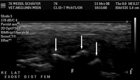
Auflagerungen, D. Schäfer, männl. 12 Jahre , Sagittalschnitt durch das Femur im Bereich der Fossa extensoria (F: Femur, ↓ Exostosen)
Stefan Perterer
"Sonographie, Röntgenologie und Pathologie des Musculus gastrocnemius im Ursprungs-bereich beim Hund"
Auflagerungen, Beagle, weibl. 10 Jahre (Nr. 26), Sagittalschnitt durch das lat. SB der li. Seite (SB: Sesambein, ↗ Ex. am Distalende des SB)
Stefan Perterer
"Sonographie, Röntgenologie und Pathologie des Musculus gastrocnemius im Ursprungs-bereich beim Hund"
Kalkschatten im Muskelbauch, D. Schäfer, männl. 9 Jahre , Sagittalschnitt durch das lat. SB und den M. gastrocnemius der re. Seite (SB: Sesambein, MG: M. gastrocnemius,
+ Kalkschatten in der Muskulatur)
Stefan Perterer
"Sonographie, Röntgenologie und Pathologie des Musculus gastrocnemius im Ursprungs-bereich beim Hund"
Kalkschatten im Muskelbauch, D. Schäfer, männl. 9 Jahre , Sagittalschnitt durch das lat. SB und den M. gastrocnemius der li. Seite (SB: Sesambein, MG: M. gastrocnemius, + Kalkschatten in der
Muskulatur)
Stefan Perterer
"Sonographie, Röntgenologie und Pathologie des Musculus gastrocnemius im Ursprungs-bereich beim Hund"
Kappenförmiger Kalkschatten, Mischling, männl. 12 Jahre , Sagittalschnitt durch das lat. SB des re. Kniegelenks (↓ Kalkschatten prox. des SB)
Stefan Perterer
"Sonographie, Röntgenologie und Pathologie des Musculus gastrocnemius im Ursprungs-bereich beim Hund"
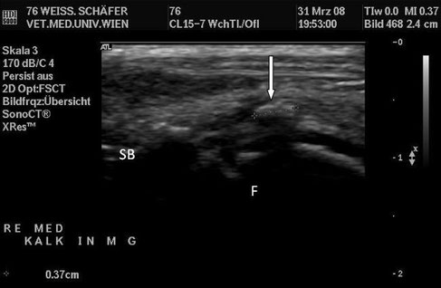
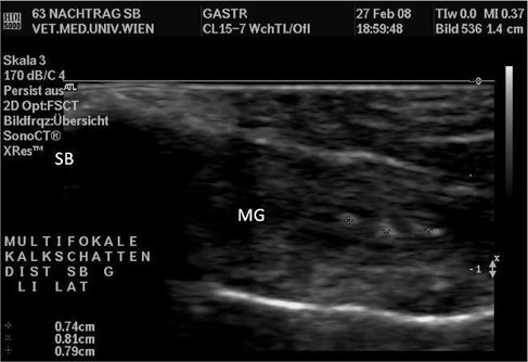
Kalkschatten distal mediales Sesambein, D. Schäfer, männl. 12 Jahre , Sagittalschnitt durch den med. Muskelbauch des M. gastrocnemius (SB: Sesambein, F: Femur, ↓
echoreiche Struktur dist. des med. SB)
Stefan Perterer
"Sonographie, Röntgenologie und Pathologie des Musculus gastrocnemius im Ursprungs-bereich beim Hund"
Exostosen, D. Schäfer, männl. 9 Jahre, Sagittalschnitt durch das laterale SB des linken Knies (SB: Sesambein, L: Leiste am kaudalen Femur, ↓ Exostosen an der Knochenleiste)
Stefan Perterer
"Sonographie, Röntgenologie und Pathologie des Musculus gastrocnemius im Ursprungs-bereich beim Hund"
Ultrasonographic examination of the left fourth rib. Irregularly thickened cortical margins associated with hypoechoic areas.
Di Tommaso M, Rocconi F, Marruchella G, et al. "Invasive pleural malignant mesothelioma with rib destruction and concurrent osteosarcoma in a dog". Acta Veterinaria Scandinavica. 2015;57:85. doi:10.1186/s13028-015-0176-1.
Diese Webseite wurde mit Jimdo erstellt! Jetzt kostenlos registrieren auf https://de.jimdo.com