

Article added / Artikel hinzugefügt 01.10.2021
Generally Articles and Discussions about Osteosarcoma in Dogs
→ Evaluations of phylogenetic proximity in a group of 67 dogs with
osteosarcoma: a pilot study
Article added / Artikel hinzugefügt 01.10.2021
Generally Articles and Discussions about Osteosarcoma in Dogs
→ Canine Periosteal Osteosarcoma
Images added / Abbildungen hinzugefügt 02.05.2019
Generally Sonography Atlas of Dogs →
Cardiovascular system → Pulmonary vessels
New subcategory added / Neue Unterkategorie hinzugefügt 02.05.2019
Generally Sonography Atlas of Dogs →
Cardiovascular system → Pulmonary vessels
Images added / Abbildungen hinzugefügt 01.05.2019
Generally Sonography Atlas of Dogs →
Cardiovascular system → Heart valvular diseases


Generally Sonography Atlas of Dogs
(Allgemeiner Sonographie-Atlas von Hunden)
Muscoskeletal System - Shoulder
(Muskoskelettales System - Schulter)
Normal shoulder: Scanning Technique & Anatomy
With special thanks to the authors of the book "Small Animal Ultrasonography" , Marc-André d’Anjou and Dominique Penninck


Diagnostic musculoskeletal shoulder ultrasound image depicting an enlargement of the supraspinatus tendon.
CANAPP, Sherman O et al. "Supraspinatus Tendinopathy in 327 Dogs: A Retrospective Study". Veterinary Evidence, [S.l.], v. 1, n. 3, jul. 2016. ISSN 2396-9776. doi:http://dx.doi.org/10.18849/ve.v1i3.32.


Diagnostic musculoskeletal shoulder ultrasound image showing a supraspinatus tendon with a mixed echogenicity, suggestive of active inflammation.
CANAPP, Sherman O et al. "Supraspinatus Tendinopathy in 327 Dogs: A Retrospective Study". Veterinary Evidence, [S.l.], v. 1, n. 3, jul. 2016. ISSN 2396-9776. doi:http://dx.doi.org/10.18849/ve.v1i3.32.


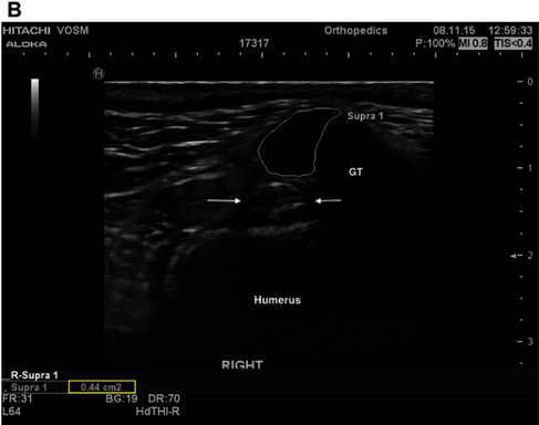
(A) A transverse ultrasonic view of the right supraspinatus tendon of patient A at the time of pre-treatment ultrasound. The supraspinatus tendon is highlighted (white outline), revealing a mottled heterogenous fiber pattern within the body of the tendon. No additional abnormalities were noted. For anatomical reference the greater tubercle (GT), humerus (humerus), and biceps tendon (arrows) are highlighted. (B) A transverse ultrasonographic view of the right supraspinatus tendon of patient A at the time of recheck ultrasound 96 days after intra-tendinous BMAC-PRP injection. The supraspinatus tendon is outlined. Note the homogenous, less echogenic (normal), fiber pattern within the body of the tendon.
McDougall Renee A., Canapp Sherman O., Canapp Debra A.: "Ultrasonographic Findings in 41 Dogs Treated with Bone Marrow Aspirate Concentrate and Platelet-Rich Plasma for a Supraspinatus Tendinopathy: A Retrospective Study"; Frontiers in Veterinary Science DOI=10.3389/fvets.2018.00098


(A) A transverse ultrasonographic view of the right supraspinatus tendon of patient B at the time of pre-treatment ultrasound, prior to an intratendinous injection with BMAC-PRP. The supraspinatus tendon is outlined. Note the generalized mottled, hyperechoic, heterogenous fiber pattern. (B) A transverse ultrasonographic view of the right supraspinatus tendon of patient B at the time of a recheck ultrasound 96 days post treatment. The supraspinatus tendon is outlined. Note the static generalized mottled, heterogenous fiber pattern. Subjectively the sonographer noted that the baseline ultrasound revealed intratendinous inflammation that had largely resolved by this time, with primarily fibrous changes remaining.
McDougall Renee A., Canapp Sherman O., Canapp Debra A.: "Ultrasonographic Findings in 41 Dogs Treated with Bone Marrow Aspirate Concentrate and Platelet-Rich Plasma for a Supraspinatus Tendinopathy: A Retrospective Study"; Frontiers in Veterinary Science DOI=10.3389/fvets.2018.00098
