Article added / Artikel hinzugefügt 01.10.2021
Generally Articles and Discussions about Osteosarcoma in Dogs
→ Evaluations of phylogenetic proximity in a group of 67 dogs with
osteosarcoma: a pilot study
Article added / Artikel hinzugefügt 01.10.2021
Generally Articles and Discussions about Osteosarcoma in Dogs
→ Canine Periosteal Osteosarcoma
Images added / Abbildungen hinzugefügt 02.05.2019
Generally Sonography Atlas of Dogs →
Cardiovascular system → Pulmonary vessels
New subcategory added / Neue Unterkategorie hinzugefügt 02.05.2019
Generally Sonography Atlas of Dogs →
Cardiovascular system → Pulmonary vessels
Images added / Abbildungen hinzugefügt 01.05.2019
Generally Sonography Atlas of Dogs →
Cardiovascular system → Heart valvular diseases


Generally Sonography Atlas of Dogs
(Allgemeiner Sonographie-Atlas von Hunden)
Cardiovascular system
(Kardiovaskuläres System)


Color Doppler window depicting the mesenteric veins.
Pablo Gomez Ochoa, Delia Lacasta, Ivan Sosa, Manuel Gascon, Juan Jose Ramos and Luis Miguel Ferrer (2011). Foundamentals and Applications of Abdominal Doppler, Ultrasound Imaging - Medical Applications, Prof. Oleg Minin (Ed.), ISBN: 978-953-307-279-1, InTech, DOI: 10.5772/20333.


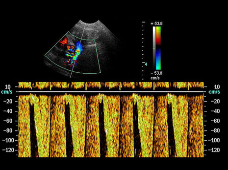
Spectral trace of an interlobar artery in a kidney
Pablo Gomez Ochoa, Delia Lacasta, Ivan Sosa, Manuel Gascon, Juan Jose Ramos and Luis Miguel Ferrer (2011). Foundamentals and Applications of Abdominal Doppler, Ultrasound Imaging - Medical Applications, Prof. Oleg Minin (Ed.), ISBN: 978-953-307-279-1, InTech, DOI: 10.5772/20333.


Continous wave Doppler in a severe aortic stenosis. A high velocity profile (6m/s) is depicted.
Pablo Gomez Ochoa, Delia Lacasta, Ivan Sosa, Manuel Gascon, Juan Jose Ramos and Luis Miguel Ferrer (2011). Foundamentals and Applications of Abdominal Doppler, Ultrasound Imaging - Medical Applications, Prof. Oleg Minin (Ed.), ISBN: 978-953-307-279-1, InTech, DOI: 10.5772/20333.


Pulsed wave Doppler in a normal portal vein. Using the sample volume the sonographer is able to define the studied volume, in this case it has a width of 4 mm.
Pablo Gomez Ochoa, Delia Lacasta, Ivan Sosa, Manuel Gascon, Juan Jose Ramos and Luis Miguel Ferrer (2011). Foundamentals and Applications of Abdominal Doppler, Ultrasound Imaging - Medical Applications, Prof. Oleg Minin (Ed.), ISBN: 978-953-307-279-1, InTech, DOI: 10.5772/20333.


Aliasing. The velocity overwhelms the máximum limit in the velocity profile (red-yellow), depicting the botom of the negative velocities (green-blue). However the blood dierction does not change.
Pablo Gomez Ochoa, Delia Lacasta, Ivan Sosa, Manuel Gascon, Juan Jose Ramos and Luis Miguel Ferrer (2011). Foundamentals and Applications of Abdominal Doppler, Ultrasound Imaging - Medical Applications, Prof. Oleg Minin (Ed.), ISBN: 978-953-307-279-1, InTech, DOI: 10.5772/20333.

Non-optimised spectral trace obtained by means of pulsed Doppler. The PRF (pulsed repetition frequency) is to low.
Pablo Gomez Ochoa, Delia Lacasta, Ivan Sosa, Manuel Gascon, Juan Jose Ramos and Luis Miguel Ferrer (2011). Foundamentals and Applications of Abdominal Doppler, Ultrasound Imaging - Medical Applications, Prof. Oleg Minin (Ed.), ISBN: 978-953-307-279-1, InTech, DOI: 10.5772/20333.


Trombus in the medial iliac artery. The blood flow is interrupted.
Pablo Gomez Ochoa, Delia Lacasta, Ivan Sosa, Manuel Gascon, Juan Jose Ramos and Luis Miguel Ferrer (2011). Foundamentals and Applications of Abdominal Doppler, Ultrasound Imaging - Medical Applications, Prof. Oleg Minin (Ed.), ISBN: 978-953-307-279-1, InTech, DOI: 10.5772/20333.


Metastasic Abdominal lymph node. The vessels could not be displayed using color Doppler, however the Power Doppler used in the image had sensitivity enough
Pablo Gomez Ochoa, Delia Lacasta, Ivan Sosa, Manuel Gascon, Juan Jose Ramos and Luis Miguel Ferrer (2011). Foundamentals and Applications of Abdominal Doppler, Ultrasound Imaging - Medical Applications, Prof. Oleg Minin (Ed.), ISBN: 978-953-307-279-1, InTech, DOI: 10.5772/20333.

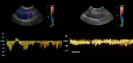
Left side of the image caudal vena cava spectral trace showing the typical bi- or tri phasic pattern. In the right side portal vein spectral trace.
Pablo Gomez Ochoa, Delia Lacasta, Ivan Sosa, Manuel Gascon, Juan Jose Ramos and Luis Miguel Ferrer (2011). Foundamentals and Applications of Abdominal Doppler, Ultrasound Imaging - Medical Applications, Prof. Oleg Minin (Ed.), ISBN: 978-953-307-279-1, InTech, DOI: 10.5772/20333.
Das gesunde Hundeherz
Mit Dank für die freundliche Genehmigung von Dr. med. vet. Ingo Schneider


With special thanks to the authors of the book "Small Animal Ultrasonography" , Marc-André d’Anjou and
Dominique Penninck

Thrombosis of the caudal vena cava. A-B: In this dog with renal adenocarcinoma, there is an echogenic thrombus filling most of the venous (CVC) lumen in transverse (A) and longitudinal (B) planes. More caudally, it extends into the right renal vein that is markedly distorted by the malignant thrombus (T). K, right kidney; RA, right adrenal gland; Ao, aorta. C-E: In this other dog with adrenal adenocarcinoma, a thrombus (between cursors) is detected in the CVC. This thrombus is associated with contrast enhancement in dorsal SPGR MR image (D) and extends into an abnormal left adrenal (Ad). The transverse T2w MR image (E) shows the connection between the malignant thrombus (T) and the left adrenal (Ad) through the phrenicoabdominal vein, just ventral to the aorta (Ao).

With special thanks to the authors of the book "Small Animal Ultrasonography" , Marc-André d’Anjou and
Dominique Penninck

Ultrasound of cardiomegaly. Outputs and dilated aorta cava.
With special thanks to Irene García Patiño (Sombra Acústica), veterinarian at the Veterinary Clinic Argos in Cee (A Coruña, Spain). http://sombraacustica.com


B-mode ultrasound image. B-mode ultrasound image showing the portal vein (PV), of one dog. Diameter measurement between callipers (0.54 cm). Transverse section at the right 11th intercostals space.
Raquel Sartor, Maria J. Mamprim, Regina F. Takahira and Mariana F. de Almeida.
"Hemodynamic evaluation of the right portal vein in healthy dogs of different body weights".
DOI: 10.1186/1751-0147-52-36


Color Doppler mapping showing the origin of the right branch of the portal vein in one dog. Note that the axis of the vessel is very close to that of the ultrasound wave, which provides an optimal insonation angle, with the flow running towards the transducer. Longitudinal section at approximately the right 10th intercostal space (RBPV: right branch of portal vein; CVC: caudal vena cava; AA: abdominal aorta; PV: portal vein).
Raquel Sartor, Maria J. Mamprim, Regina F. Takahira and Mariana F. de Almeida.
"Hemodynamic evaluation of the right portal vein in healthy dogs of different body weights".
DOI: 10.1186/1751-0147-52-36

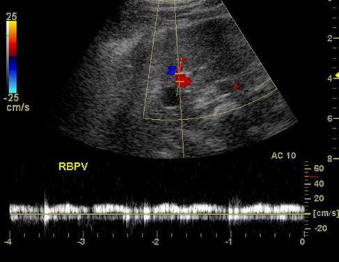
Spectral Doppler mapping of the right branch of the portal vein (RBPV) of one dog. Longitudinal section at approximately the right 10th intercostal space. Note the insonation angle (12 degrees), which provides measurements with minimum margin of error. The flow is monophasic and presents low pulsatility.
Raquel Sartor, Maria J. Mamprim, Regina F. Takahira and Mariana F. de Almeida.
"Hemodynamic evaluation of the right portal vein in healthy dogs of different body weights".
DOI: 10.1186/1751-0147-52-36