

Article added / Artikel hinzugefügt 01.10.2021
Generally Articles and Discussions about Osteosarcoma in Dogs
→ Evaluations of phylogenetic proximity in a group of 67 dogs with
osteosarcoma: a pilot study
Article added / Artikel hinzugefügt 01.10.2021
Generally Articles and Discussions about Osteosarcoma in Dogs
→ Canine Periosteal Osteosarcoma
Images added / Abbildungen hinzugefügt 02.05.2019
Generally Sonography Atlas of Dogs →
Cardiovascular system → Pulmonary vessels
New subcategory added / Neue Unterkategorie hinzugefügt 02.05.2019
Generally Sonography Atlas of Dogs →
Cardiovascular system → Pulmonary vessels
Images added / Abbildungen hinzugefügt 01.05.2019
Generally Sonography Atlas of Dogs →
Cardiovascular system → Heart valvular diseases


Generally Sonography Atlas of Dogs
(Allgemeiner Sonographie-Atlas von Hunden)
Muscoskeletal System - Hock
(Muskoskelettales System - Sprunggelenk)


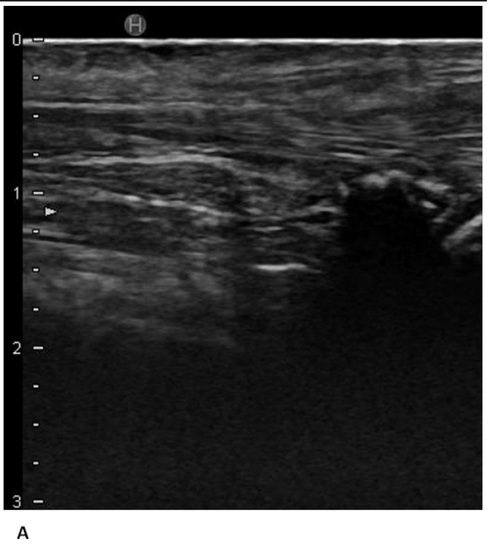
Grade I Strain
A- Longitudinal view of grade I strain of common tendon.
Evidence of right CT early disruption with mild hyperechoic/hypoechoic foci noted at the point of insertion.
B- Longitudinal view of grade I strain of common and gastrocnemius tendons.
There was a generalised hypoechoic fibre pattern to the CT and GT with no disruption noted. In addition the common tendon bursa also showed chronic hyperechoic changes consistent with
bursitis.
GAMBLE, Lauri-Jo; CANAPP, Debra A; CANAPP, Sherman O. Evaluation of Achilles Tendon Injuries with Findings from Diagnostic Musculoskeletal Ultrasound in Canines – 43 Cases. Veterinary Evidence, [S.l.], v. 2, n. 3, sep. 2017. ISSN 2396-9776.


Grade II Strain
Longitudinal view grade II strain gastrocnemius and common tendons with grade I superficial digital flexor tendon.
The right SDFT appeared slightly thickened but showed overall good pattern. There was a mild amount of periosteal hyperechoic changes noted at the deep margin of the calcaneus. There was
moderate disruption of the GT, mid tendon with normal identifiable pattern at the distal 1/3 of the tendon and more disruption note proximal to that point. The CT was moderately disrupted at the
point of insertion with severe hypoechoic fib pattern and hyperechoic foci at the point of insertion at the calcaneus. In addition there were moderate areas of anechoic findings, indicating
moderate fluid accumulation within the bursa.
GAMBLE, Lauri-Jo; CANAPP, Debra A; CANAPP, Sherman O. Evaluation of Achilles Tendon Injuries with Findings from Diagnostic Musculoskeletal Ultrasound in Canines – 43 Cases. Veterinary Evidence, [S.l.], v. 2, n. 3, sep. 2017. ISSN 2396-9776.

Grade III Strain
A- Longitudinal view of a grade III strain of the Achilles tendon (all components involved).
This Achilles tendon was hypoechoic with loss of normal and mottled appearance. There was a significant amount of hypoechoic soft tissue changes with lack of fibre pattern noted
superficially at and just proximal to the calcaneus. The calcaneal tuberosity also had visible periosteal reaction.
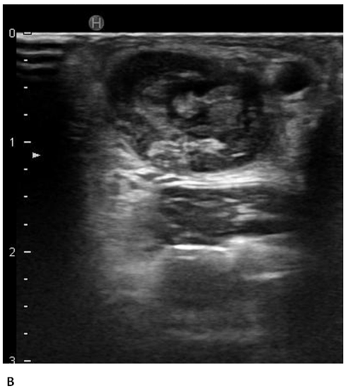
B- Cross-sectional view of a grade III strain of the Achilles tendon (all components involved).
There was severe disruption of the GT with a minor amount of identifiable fibre pattern at the distal 1/3 of the tendon, attaching to the calcaneus. There were also age/degenerative hyperechoic
changes noted within the common tendon bursa. In general the areas of loss of fibre pattern showed hypoechoic tissue with some fibrous tissue.
GAMBLE, Lauri-Jo; CANAPP, Debra A; CANAPP, Sherman O. Evaluation of Achilles Tendon Injuries with Findings from Diagnostic Musculoskeletal Ultrasound in Canines – 43 Cases. Veterinary Evidence, [S.l.], v. 2, n. 3, sep. 2017. ISSN 2396-9776.

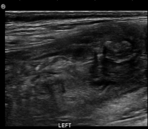
Alteration of the Musculotendinous Junction (MTJ)
Longitudinal view of the musculotendinous junction.
The musculotendinous junction of both the GT and the SDF were significantly altered with lack of normal muscle of fibre tendon. Several hyperechoic fibs were ‘’coiled up’’ amongst the altered
muscle tissue which may be representing retracted tendon fibs from a distal rupture in addition to a MTJ rupture.
GAMBLE, Lauri-Jo; CANAPP, Debra A; CANAPP, Sherman O. Evaluation of Achilles Tendon Injuries with Findings from Diagnostic Musculoskeletal Ultrasound in Canines – 43 Cases. Veterinary Evidence, [S.l.], v. 2, n. 3, sep. 2017. ISSN 2396-9776.


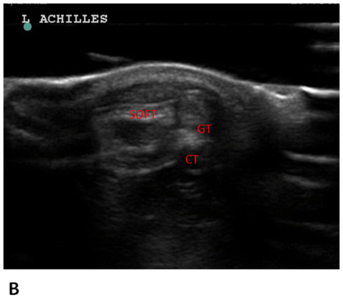


Figure A: Normal Achilles Longitudinal and Cross-Sectional Views
Longitudinal view of a normal Achilles tendon.
In longitudinal images of the distal part of the tendon the gastrocnemius tendon (GT) lies between the superficial digital flexor tendon (SDFT) and the common tendon (CT). The insertional bursa
(Bursa) of the common tendon is visible at proximity of the calcaneal tuberosity (Ca) and its acoustic shadowing.
Figure B: Cross-sectional view of a normal Achilles tendon - proximal
In cross-sectional view, the appearance of the 3 components of the Achilles tendon remains virtually unchanged in the proximal and middle parts of the tendon.
description of image here
Figure 6C: Cross-sectional view of a normal Achilles tendon - distal.
Toward the distal part, the superficial digital flexor tendon (SDFT) moves to a more superficial position.
Figure 6D: Cross-sectional view of the SDFT.
Visualization of the SDFT as it courses over the calcaneal tuberosity (Ca).
GAMBLE, Lauri-Jo; CANAPP, Debra A; CANAPP, Sherman O. Evaluation of Achilles Tendon Injuries with Findings from Diagnostic Musculoskeletal Ultrasound in Canines – 43 Cases. Veterinary Evidence, [S.l.], v. 2, n. 3, sep. 2017. ISSN 2396-9776.
