

Article added / Artikel hinzugefügt 01.10.2021
Generally Articles and Discussions about Osteosarcoma in Dogs
→ Evaluations of phylogenetic proximity in a group of 67 dogs with
osteosarcoma: a pilot study
Article added / Artikel hinzugefügt 01.10.2021
Generally Articles and Discussions about Osteosarcoma in Dogs
→ Canine Periosteal Osteosarcoma
Images added / Abbildungen hinzugefügt 02.05.2019
Generally Sonography Atlas of Dogs →
Cardiovascular system → Pulmonary vessels
New subcategory added / Neue Unterkategorie hinzugefügt 02.05.2019
Generally Sonography Atlas of Dogs →
Cardiovascular system → Pulmonary vessels
Images added / Abbildungen hinzugefügt 01.05.2019
Generally Sonography Atlas of Dogs →
Cardiovascular system → Heart valvular diseases


Generally Sonography Atlas of Dogs - Genitourinary system
(Allgemeiner Sonographie-Atlas von Hunden) - (Urogenitales System)
Pregnancy
(Schwangerschaft)


Gestation. VE = vesicle embryonic ;
RP = rear reinforcement .
N. Díez Bru
"Ecografía abdominal en pequeños animales."
CLINICA VETERINARIA DE PEQUEÑOS ANIMALES
Volumen 12, Número 3, Julio/Septiembre 1992


Dog pregnancy sonographic picture
(from http://idhumanbody.com)


Dogs embryo - 37 days
(from http://idhumanbody.com)


Ultrasonograms of the extra-fetal structures in pregnant Miniature Schnauzer bitches. Day 18: Transverse image of the first detection of an anechoic gestational sac. Day 21: Longitudinal image of the gestational sac. An echogenic inner placental layer was detected in the uterine wall. Day 27: Longitudinal image of the gestational sac contained an embryo and the tubular shape of the yolk sac membrane (white arrows). The zonary placenta (white arrowheads) was cylindrical in shape and appeared folded inward at the edges. Day 28: The amnionic membrane (white arrows) appeared faint.
30 September 2007
2868137
Journal of Veterinary Science
The Korean Society of Veterinary Science


Ultrasonograms of the fetal structures in pregnant Miniature Schnauzer bitches. Day 23: Transverse image of the gestational sac contained an oblong-shaped embryo (white arrowhead) apposed to the uterine wall. Day 32: Longitudinal image of a fetus with an anechoic area in the head and forelimb buds (white arrowheads). The fetal crown-rump length was 20 mm. Day 38: Longitudinal image of a fetus with hyperechoic skeletal structures in the head and thoracic wall (white arrowheads). Day 45: Longitudinal image of a fetal vertebral column and kidney (white arrowheads).
30 September 2007
2868137
Journal of Veterinary Science
The Korean Society of Veterinary Science

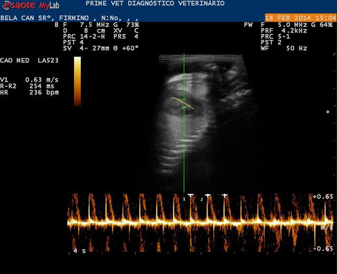
Farbdoppler fetale Zirkulation
Mit Dank für die freundliche Genehmigung an Dr. Nitsch http://www.drnitsch.de/


Gerät mit einem PW Doppler. Die Sonde liegt auf dem Herzen eines Hundewelpen. Der Herzschlag des Welpen ist über Lautsprecher zu hören und auch grafisch dargestellt (die Zacken auf der unteren Linie). So kann man bei einer Geburt beurteilen, ob es dem Welpen in der Mutter gut geht.
Mit freundlicher Genehmigung der Tierarztpraxis Dr. Peter Neu, Coburg.

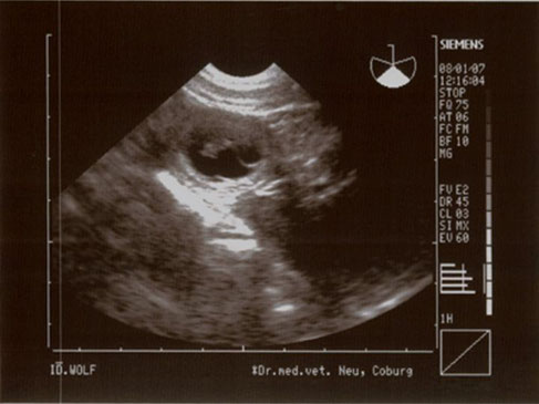

Nachweis der Trächtigkeit. Sie ist beim Hund etwa vom 21.Tag möglich.
In diesem Stadium sieht man die Foeten als „Fruchtampullen“ und kann recht gut ihre Zahl bestimmen. In einem späteren Stadium, kann man lebenswichtige Organe wie Herz, Leber und Magen der Foeten sehen. Herzschlag und Fruchtbewegungen lassen sich schön zeigen.
Mit freundlicher Genehmigung der Tierarztpraxis Dr. Peter Neu, Coburg.


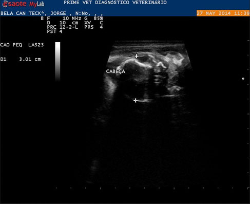
Bitch 32 days of gestation , there is more apparent vesicles, but with training heart and some organs.
With special thanks to Priscilla Pinel, Medical Veterinary.
Currently serves in veterinary clinics and homes for the municipality of Rio de Janeiro ( south, north and west ).
http://veterinariapriscillapinel.com.br


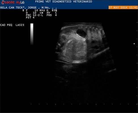
Images of another bitch in 43 days, presence of bone formation and more apparent internal organs.
With special thanks to Priscilla Pinel, Medical Veterinary.
Currently serves in veterinary clinics and homes for the municipality of Rio de Janeiro ( south, north and west ).
http://veterinariapriscillapinel.com.br


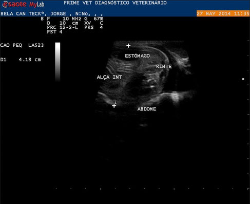
Images of another bitch in 58 days, fully formed internal organs, with the presence of intestinal motility , kidneys view, lung and liver.
With special thanks to Priscilla Pinel, Medical Veterinary.
Currently serves in veterinary clinics and homes for the municipality of Rio de Janeiro ( south, north and west ).
http://veterinariapriscillapinel.com.br


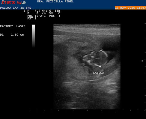
Images of a bitch now 65 days.
With special thanks to Priscilla Pinel, Medical Veterinary.
Currently serves in veterinary clinics and homes for the municipality of Rio de Janeiro ( south, north and west ).
http://veterinariapriscillapinel.com.br


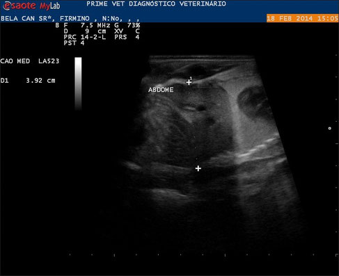
Pregnancy , embryonic absorption and fetal death.
Bitch pregnant with approximately 45 days.

Same animal in which the right horn has two gestacionárias vesicles without the presence of fetal heartbeat and embryo disintegration.
With special thanks to Priscilla Pinel, Medical Veterinary.
Currently serves in veterinary clinics and homes for the municipality of Rio de Janeiro ( south, north and west ).
http://veterinariapriscillapinel.com.br


Fetal death likely hemoparasitosis
3 years bitch crossed to 35 days, with bloody
discharge.
In ultrasonographic examination were viewed at least six gestational vesicles with
the presence of fetuses without beating with thickening of the gallbladder wall, and
having at least one well-formed fetus with definition of chest to abdomen, and
the other with bone formation and little definition thorax and abdomen.
It was still seen good hypoechoic enlarged spleen and liver bitch.
Suggesting a possible hemoparasitosis and fetal death at various gestational
stages.
In some cases hemoparasitoses (tick disease), end up very lowering
the immune system, and this pregnancy is interrupted, or there may be
birth defects. The hypoechoic liver also justify an acute liver disease
(toxemia).
With special thanks to Priscilla Pinel, Medical Veterinary.
Currently serves in veterinary clinics and homes for the municipality of Rio de Janeiro ( south, north and west ).
http://veterinariapriscillapinel.com.br


Planimetric image of the ultrasound beam’s action angle in the placenta ring. The hachured area represents the angle (approximately ) of the beam’s incidence on the placenta ring pars intermedia, the point where thickness measurements were performed (a). (b) Corresponding ultrasound image.
André Luiz Louzada Maldonado, Edward Araujo Júnior, Débora Sartori Mendonça, Luciano Marcondes Machado Nardozza, Antonio Fernandes Moron, and Sérgio Aron Ajzen, “Ultrasound Determination of Gestational Age Using Placental Thickness in Female Dogs: An Experimental Study,” Veterinary Medicine International, vol. 2012, Article ID 850867, 6 pages, 2012. doi:10.1155/2012/850867


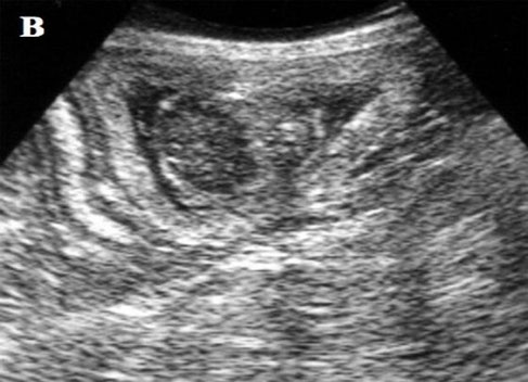
Ultrasonograms of the fetal structures in pregnant bitches.
Day 16: Transverse image of the first detection of a gestational sac (GS: gestational sac).
Day 24: Longitudinal image of the gestational sac contained an embryo the tubular shape of the yolk sac membrane (A: amnios, B: bladder, GS: gestational sac).
Day 36: Longitudinal image of a fetus (B: bladder, F: liver, L: lung, H: heart).
Aissi, A.: "Aspects of Ultrasonographic Diagnostics of Pregnancy in Bitches depending on the first mating"; Veterinary World, Vol.1(10): 293-295
