

Article added / Artikel hinzugefügt 01.10.2021
Generally Articles and Discussions about Osteosarcoma in Dogs
→ Evaluations of phylogenetic proximity in a group of 67 dogs with
osteosarcoma: a pilot study
Article added / Artikel hinzugefügt 01.10.2021
Generally Articles and Discussions about Osteosarcoma in Dogs
→ Canine Periosteal Osteosarcoma
Images added / Abbildungen hinzugefügt 02.05.2019
Generally Sonography Atlas of Dogs →
Cardiovascular system → Pulmonary vessels
New subcategory added / Neue Unterkategorie hinzugefügt 02.05.2019
Generally Sonography Atlas of Dogs →
Cardiovascular system → Pulmonary vessels
Images added / Abbildungen hinzugefügt 01.05.2019
Generally Sonography Atlas of Dogs →
Cardiovascular system → Heart valvular diseases


Generally Sonography Atlas of Dogs - Genitourinary system
(Allgemeiner Sonographie-Atlas von Hunden) - (Urogenitales System)
Male reproduktion
(Männliche Fortpflanzung)


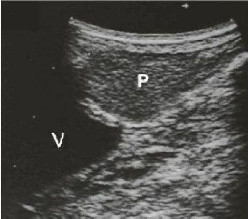
Intraprostlitico cyst ( Q ) ; P = prostate ; V = bladder neck .
N. Díez Bru
"Ecografía abdominal en pequeños animales."
CLINICA VETERINARIA DE PEQUEÑOS ANIMALES
Volumen 12, Número 3, Julio/Septiembre 1992


Ultrasound image of canine reproduction track image of dogs testical
(from http://idhumanbody.com)


Ultrasonographic and colour doppler images of right testis: the normal echographic pattern of testis is not recognizable. The mediastinum testis is not showed and blood flows is visible within the parenchyma.
Ultrasonographic image of left testis: The testis is small and iperechoic. None Doppler signal are present and it appears as fat tissue.
From: Bigliardi et al: Clinical, genetic and pathological features of male pseudohermaphroditism in dog


Figure 1. Prostate carcinoma: increase of volume and change of normal echographic findings.
Figure 2. Prostate carcinoma: in the bottom of the image is present one para-prostatic cyst (white arrow). Note the increase of volume and alteration of prostate tissue.
Figure 3. Prostate carcinoma: Neutered dog. The structure of prostate shows ipoechoic (white arrow) and anechoic areas (red arrow).
("Canine Prostate Carcinoma: Four Clinical Cases in Sexually Intact and Neutered Dogs", DOI:10.4236/oju.2012.24042, from: http://file.scirp.org)


Hund mit abdominalem Kryptorchismus und Aszites (links: Hoden, rechts: Nebenhoden)
http://de.wikipedia.org/wiki/Hoden
„Abdominaler Kryptorchismus Sono“. Lizenziert unter CC BY 2.5 über Wikimedia Commons - http://commons.wikimedia.org/wiki/File:Abdominaler_Kryptorchismus_Sono.jpg#mediaviewer/File:Abdominaler_Kryptorchismus_Sono.jpg


Transitional cell carcinoma and regional lymphadenopathy in an old, neutered dog with hematuria and dysuria. Sagittal (A) and transverse (B) images of the bladder neck and prostate. Strongly shadowing hyperechoic foci are in the prostate, and a soft-tissue projection is in the bladder lumen (arrow). Sagittal images of right medial iliac (C) and hypogastric (D) lymph nodes, which appear enlarged and irregular. The medial iliac node (C) is relatively uniform and nearly anechoic in comparison with the hypogastric (D) node that is heterogeneous and coarse in echotexture. These lymph nodes are adjacent to the external and internal iliac vessels, respectively.
With special thanks to the authors of the book "Small Animal Ultrasonography" , Marc-André d’Anjou and Dominique Penninck


Sonographische Darstellung einer durch BPH bedingten Koprostase beim Hund
http://de.wikipedia.org/wiki/Benigne_Prostatahyperplasie
„Prostatahyperplasie Koprostase Sono“ von Kalumet - Eigenes Werk. Lizenziert unter GFDL über Wikimedia Commons - http://commons.wikimedia.org


Typical Doppler ultrasound waveforms from the testicular artery of post-pubertal dogs. Images were recorded from (A) distal supra-testicular artery, (B) marginal artery, (C) intra-testicular artery.
"Regional differences of testicular artery blood flow in post pubertal and pre-pubertal dogs", Mírley B de Souza, Claudia C Barbosa, Gary CW England, Antonio C Mota Filho, Carmen Vládia S Sousa, Gabriela G de Carvalho, Herlon Victor R Silva, José N Pinto, Jussiara CS Linhares and Lúcia DM Silva
doi:10.1186/s12917-015-0363-3


Typical Doppler ultrasound waveforms from the testicular artery of pre-pubertal dogs. Images were recorded from (A) distal supra-testicular artery, (B) marginal artery, (C) intra-testicular artery.
"Regional differences of testicular artery blood flow in post pubertal and pre-pubertal dogs", Mírley B de Souza, Claudia C Barbosa, Gary CW England, Antonio C Mota Filho, Carmen Vládia S Sousa, Gabriela G de Carvalho, Herlon Victor R Silva, José N Pinto, Jussiara CS Linhares and Lúcia DM Silva
doi:10.1186/s12917-015-0363-3

Prostatic image in a dog using transrectal ultrasound.
J Thibaut *, J Santander, M Mieres
"Comparative study of the canine prostate using transrectal and transabdominal ultrasonographic techniques"
http://dx.doi.org/10.4067/S0301-732X2009000100008


Prostatic image in a dog using transrectal ultrasound.
J Thibaut *, J Santander, M Mieres
"Comparative study of the canine prostate using transrectal and transabdominal ultrasonographic techniques"
http://dx.doi.org/10.4067/S0301-732X2009000100008


Ectopic testes in a canine Fox
Paulistinha 9 months.
With special thanks to Priscilla Pinel, Medical Veterinary.
Currently serves in veterinary clinics and homes for the municipality of Rio de Janeiro ( south, north and west ).
http://veterinariapriscillapinel.com.br


Ectopic left testicle of a canine Australian Red 10 month.
With special thanks to Priscilla Pinel, Medical Veterinary.
Currently serves in veterinary clinics and homes for the municipality of Rio de Janeiro ( south, north and west ).
http://veterinariapriscillapinel.com.br


Echography showed a parenchyma of a heterogeneous texture and presence of several cavities prostate sufferers hypoechogenes with a normaly vesical and renal. P: Prostate; PK: Prostatic kystic; B: Normal blader.
Ahmed Boucif, Khadidja Madani, Aboud Boulkaboul and Khaled Slimani
"Chronic Prostatitis (CP) in Atlas Shepherd Dog: A Case-Control Study"
doi: 10.4172/2327-5073.1000197


Testicular atrophy in an 1.5-year-old, right sided cryptorchid Shi Tzu. The right testicle is found in the abdominal cavity (A), adjacent to intestinal loops. It is small (1.4 cm long) while the left scrotal testicle (B, between cursors) is normal in size (2.2 cm long).
With special thanks to the authors of the book "Small Animal Ultrasonography" , Marc-André d’Anjou and Dominique Penninck


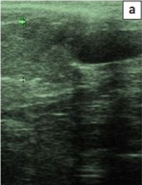
Ultrasound through the scrotum.
In the picture we see on the right testicle normal echotexture. On
the left side is the other testicle with a normal area echotexture but another
with mixed picture.
With special thanks to Irene García Patiño (Sombra Acústica), veterinarian at the Veterinary Clinic Argos in Cee (A Coruña, Spain). http://sombraacustica.com


Scrotal hernia. In both ultrasounds seen within the scrotal sac, bowel loops left and right to the testicle. In the testis is a central hyperechoic line corresponding to the mediastinum testis.
With special thanks to Irene García Patiño (Sombra Acústica), veterinarian at the Veterinary Clinic Argos in Cee (A Coruña, Spain). http://sombraacustica.com


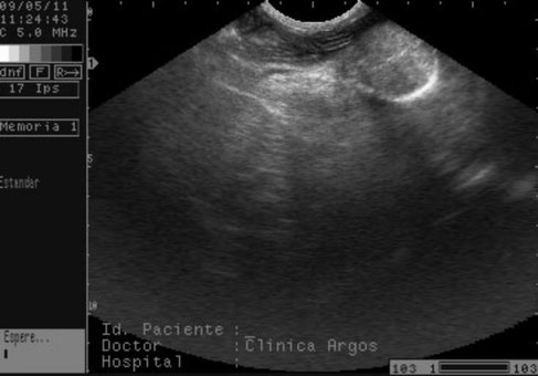
Prostatic cyst.
With special thanks to Irene García Patiño (Sombra Acústica), veterinarian at the Veterinary Clinic Argos in Cee (A Coruña, Spain). http://sombraacustica.com


Sagittal diagnostic ultrasound images of the caudal os penis region (a) the obstructive haemangiosarcoma mass between the cursors is located cranially to the anechoic dilated urethra and (b) slightly different imaging plane showing the irregular hyperechoic os penis remnants with some acoustic shadowing within the haemangiosarcoma.
Richard K. Burchell, Robert M. Kirberger,
Drienie D. (Didi) Janse van Rensburg, "Haemangiosarcoma of the os penis in a dog: The most common neoplasm of the canine penis", http://dx.doi.org/10.4102/
jsava.v85i1.1092


Screenshot der 3D-Software
Ausgangsposition für das longitudinale
Durchmustern des Würfels
Longitudinales Durchmustern des
Würfels
Ausgangsposition für das transversale
Durchmustern des Würfels
Transversales Durchmustern des Würfels
„Manuelle Planimetrie": Markieren
des Umfangs der Prostata
Kerstin Huemer, "VOLUMETRIE DER
PROSTATA DES RÜDEN MIT DER
3D-SONOGRAPHIE-SOFTWARE 3DVET",
Dezember 2008


(a) Transverse ultrasound image of case 2 showing severely dilated small intestinal loop with partial loss of wall layering and increased overall echogenicity of the intestinal wall. Next to the dilated loop there is normal looking small intestine. (b) Longitudinal image of an affected small intestinal loop.
Elina Rautala, Pia Björkenheim, Merja Laitinen: "Radiographic and Ultrasonographic Findings in Three Surgically Confirmed Cases of Small Intestinal Ischemia Related to Mesenteric Volvulus
or Intestinal Torsion in Dogs";
DOI: 10.4236/ojvm.2017.79010


(a) Transverse image of the affected small intestinal loop of case 3 showing no signal on Doppler; (b) Compared with normal looking adjacent segment of small intestine showing clear Doppler signal.
Elina Rautala, Pia Björkenheim, Merja Laitinen: "Radiographic and Ultrasonographic Findings in Three Surgically Confirmed Cases of Small Intestinal Ischemia Related to Mesenteric Volvulus
or Intestinal Torsion in Dogs";
DOI: 10.4236/ojvm.2017.79010.


Longitudinal image of the affected small intestine showing small gas bubbles within or adjacent to the wall of the small intestinal segment and some strand-like structures of soft tissue echogenicity in the lumen of the intestine.
Elina Rautala, Pia Björkenheim, Merja Laitinen: "Radiographic and Ultrasonographic Findings in Three Surgically Confirmed Cases of Small Intestinal Ischemia Related to Mesenteric Volvulus
or Intestinal Torsion in Dogs";
DOI: 10.4236/ojvm.2017.79010.
