

Article added / Artikel hinzugefügt 01.10.2021
Generally Articles and Discussions about Osteosarcoma in Dogs
→ Evaluations of phylogenetic proximity in a group of 67 dogs with
osteosarcoma: a pilot study
Article added / Artikel hinzugefügt 01.10.2021
Generally Articles and Discussions about Osteosarcoma in Dogs
→ Canine Periosteal Osteosarcoma
Images added / Abbildungen hinzugefügt 02.05.2019
Generally Sonography Atlas of Dogs →
Cardiovascular system → Pulmonary vessels
New subcategory added / Neue Unterkategorie hinzugefügt 02.05.2019
Generally Sonography Atlas of Dogs →
Cardiovascular system → Pulmonary vessels
Images added / Abbildungen hinzugefügt 01.05.2019
Generally Sonography Atlas of Dogs →
Cardiovascular system → Heart valvular diseases


Generally Sonography Atlas of Dogs - Genitourinary system
(Allgemeiner Sonographie-Atlas von Hunden) - (Urogenitales System)
Kidney Page 2
(Nieren Seite 2)

Ultrasonographic appearance (2D) of right kidney in midsagittal plane with “end-stage” kidney disease in 4-year-old, spayed, female, Boxer dog with increased medullary echogenicity.
Kumar V, Kumar A, Varshney AC. "Ultrasonographic Imaging for Structural Characterization of Renal Affections and Diagnosis of Associated Chronic Renal Failure in 10 Dogs". ISRN Veterinary Science. 2011;2011:901713. doi:10.5402/2011/901713.


2D ultrasonogram in sagittal plane of right kidney in a 9-year-old, intact male, Gaddi cross dog revealing renal pelvic dilatation (pyelectasia) and atrophy of renal medulla.
Kumar V, Kumar A, Varshney AC. "Ultrasonographic Imaging for Structural Characterization of Renal Affections and Diagnosis of Associated Chronic Renal Failure in 10 Dogs". ISRN Veterinary Science. 2011;2011:901713. doi:10.5402/2011/901713.

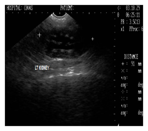
Ultrasonographic appearance (2D) of left kidney in sagittal plane showing marked renomegaly with renal pelvic dilatation and hyperechoic medullary rim in a 9-year-old, intact male, Gaddi cross dog.
Kumar V, Kumar A, Varshney AC. "Ultrasonographic Imaging for Structural Characterization of Renal Affections and Diagnosis of Associated Chronic Renal Failure in 10 Dogs". ISRN Veterinary Science. 2011;2011:901713. doi:10.5402/2011/901713.

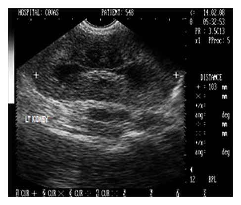
2D sonogram in midsagittal scan of left kidney in a two-year-old, intact male, German shepherd dog with ultrasonographic impression of an enlarged kidney measuring 103 mm in length and revealing increased cortical and medullary echogenicity.
Kumar V, Kumar A, Varshney AC. "Ultrasonographic Imaging for Structural Characterization of Renal Affections and Diagnosis of Associated Chronic Renal Failure in 10 Dogs". ISRN Veterinary Science. 2011;2011:901713. doi:10.5402/2011/901713.


Ultrasonographic appearance (2D) in transverse scan of the enlarged right kidney with renal width measuring 62 mm and hyperechoic renal cortex in a two-year-old, intact male, German shepherd dog.
Kumar V, Kumar A, Varshney AC. "Ultrasonographic Imaging for Structural Characterization of Renal Affections and Diagnosis of Associated Chronic Renal Failure in 10 Dogs". ISRN Veterinary Science. 2011;2011:901713. doi:10.5402/2011/901713.


2D left renal sonogram in sagittal plane with increased cortical echogenicity and renal length of 61 mm in a 17-year-old, 6 kg body weight, intact female, Pomeranian dog.
Kumar V, Kumar A, Varshney AC. "Ultrasonographic Imaging for Structural Characterization of Renal Affections and Diagnosis of Associated Chronic Renal Failure in 10 Dogs". ISRN Veterinary Science. 2011;2011:901713. doi:10.5402/2011/901713.


B+B sonograph of left and right kidneys with increased cortical echogenicities in a 17-year-old, intact female, Pomeranian dog.
Kumar V, Kumar A, Varshney AC. "Ultrasonographic Imaging for Structural Characterization of Renal Affections and Diagnosis of Associated Chronic Renal Failure in 10 Dogs". ISRN Veterinary Science. 2011;2011:901713. doi:10.5402/2011/901713.


(a) 2D sonogram of right kidney in midsagittal scan revealing large hyperechoic structure in the renal pelvis with intense distal acoustic shadowing and masking of the far calculi renal cortex in a 9-year-old, intact male, Doberman pinscher dog. (b) 2D sonograph (transverse scan) of right kidney revealing scattered hyperechoic densities with acoustic shadowing suggesting renal medullary mineralization in a six-year-old, intact female, mixed breed dog.
Kumar V, Kumar A, Varshney AC. "Ultrasonographic Imaging for Structural Characterization of Renal Affections and Diagnosis of Associated Chronic Renal Failure in 10 Dogs". ISRN Veterinary Science. 2011;2011:901713. doi:10.5402/2011/901713.


Right kidney sonogram (2D) in sagittal plane revealing two spherical cortical cystic lesions with anechoic fluid in a 16-year-old, neutered male, mixed breed dog.
Kumar V, Kumar A, Varshney AC. "Ultrasonographic Imaging for Structural Characterization of Renal Affections and Diagnosis of Associated Chronic Renal Failure in 10 Dogs". ISRN Veterinary Science. 2011;2011:901713. doi:10.5402/2011/901713.

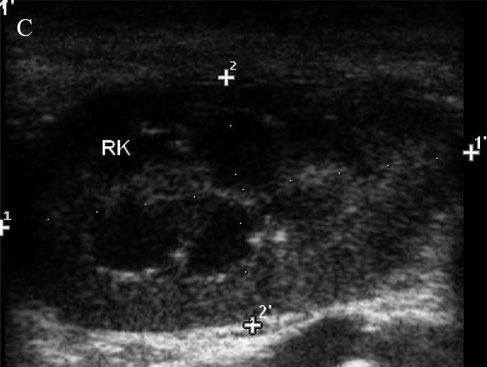
Sagittal view of the right kidney (RK). Cursors represent the length (11’) and the width (22’) of RK.
WICKRAMASEKARA RAJAPAKSHAGE, B. K., ELLEARAEWE GARUHAMILAGE, J. P. K., DE SILVA, D. D. N., & DANGOLLA, A. (2016). "Dimensional ultrasonographic relationship of the right lobe of pancreas with associated anatomic landmarks in clinically normal dogs". http://doi.org/10.1292/jvms.15-0209


CEUS renal perfusion images. Q-LAB quantification software was used for the plotting Time Intensity Curves (TIC) which was generated from the Region of Interest (ROI).
Xiu Xia Fang, Bing Hui Fan, Ai-Qing Zhao, Ping Ping and Cui Yan, "Quantitative analysis of contrast-enhanced ultrasound in the dog's acute renal failure", Biomedical Research (2017) Volume 28, Issue 16, ISSN 0970-938X


LONGITUDINAL SECTION OF A KIDNEY OBTAINED USING ULTRASONOGRAPHY
Barrera, Rafael; Duque, Javier; Ruiz, Patricia; Zaragoza, Concepción: "ACCURACY OF ULTRASONOGRAPHIC MEASUREMENTS OF KIDNEY DOG FOR CLINICAL USE",
Revista Científica, vol. XIX, núm. 6, noviembre-diciembre, 2009, pp. 576-583


CORONAL SECTION OF A KIDNEY OBTAINED USING ULTRASONOGRAPHY.
Barrera, Rafael; Duque, Javier; Ruiz, Patricia; Zaragoza, Concepción: "ACCURACY OF ULTRASONOGRAPHIC MEASUREMENTS OF KIDNEY DOG FOR CLINICAL USE",
Revista Científica, vol. XIX, núm. 6, noviembre-diciembre, 2009, pp. 576-583


Nephritis: (a) In two-dimensional
ultrasonogram of kidney, the echogenicity of renal parenchyma has increased without differentiation of cortex and medulla (red arrow). Echogenic renal crest (yellow
arrow), (b) in three-dimensional (3D) renal parenchyma without distinction of cortex and medulla (red arrow). Renal crest visible in the center (black arrow).
Dinesh Dehmiwal, S. M. Behl, Prem Singh, Rishi Tayal, Madan Pal and R. K. Chandolia: "Diagnosis of pathological conditions of kidney by two-dimensional and
three-dimensional ultrasonographic imaging in dogs"; Veterinary World, EISSN: 2231-0916


Nephritis: (a) In two-dimensional
ultrasonogram of kidney, the echogenic of renal diverticulae (red arrow), renal crest (black arrow) and corticomedullary
junction (blue), (b) in three-dimensional ultrasonogram of kidney, homogeneous without differentiation of medulla (red arrow). Echogenic renal sinus in the center (yellow arrow).
Dinesh Dehmiwal, S. M. Behl, Prem Singh, Rishi Tayal, Madan Pal and R. K. Chandolia: "Diagnosis of pathological conditions of kidney by two-dimensional and
three-dimensional ultrasonographic imaging in dogs"; Veterinary World, EISSN: 2231-0916


End-stage kidney: (a) In two-dimensional ultrasonogram of kidney, small sized kidney appears without distinct margins (red arrow)
and differentiation of cortex and medulla, (b) in three-dimensional ultrasonogram of kidney, small sized kidney appears without distinct margins (red arrow).
Dinesh Dehmiwal, S. M. Behl, Prem Singh, Rishi Tayal, Madan Pal and R. K. Chandolia: "Diagnosis of pathological conditions of kidney by two-dimensional and
three-dimensional ultrasonographic imaging in dogs"; Veterinary World, EISSN: 2231-0916


Hydronephrosis: (a) In two-dimensional
ultrasonogram of kidney with dilated renal pelvis (red arrow). Atrophied renal parenchyma with loss of architectural details (black arrow), (b) in three-dimensional
ultrasonogram dilated renal pelvis (red arrow).
Dinesh Dehmiwal, S. M. Behl, Prem Singh, Rishi Tayal, Madan Pal and R. K. Chandolia: "Diagnosis of pathological conditions of kidney by two-dimensional and
three-dimensional ultrasonographic imaging in dogs"; Veterinary World, EISSN: 2231-0916


Hydronephrosis: (a) In two-dimensional
ultrasonogram of kidney with dilated renal pelvis (red arrow). Atrophied renal parenchyma with loss of architectural details (black arrow), (b) in three-dimensional
ultrasonogram fluid filled areas (red arrow), course of ureter (yellow arrow) and ascetic fluid outsite the kidney (green arrow).
Dinesh Dehmiwal, S. M. Behl, Prem Singh, Rishi Tayal, Madan Pal and R. K. Chandolia: "Diagnosis of pathological conditions of kidney by two-dimensional and
three-dimensional ultrasonographic imaging in dogs"; Veterinary World, EISSN: 2231-0916


Nephrolithiasis: (a) In two-dimensional
ultrasonogram of kidney the nephrolith is clearly visible (red arrow). The architectural details showing renal crest (black arrow) and divarticulae (green arrow), (b) in
three-
dimensional ultrasonogram of kidney the nephrolith (red arrow) and renal parenchyma (black arrow).
Dinesh Dehmiwal, S. M. Behl, Prem Singh, Rishi Tayal, Madan Pal and R. K. Chandolia: "Diagnosis of pathological conditions of kidney by two-dimensional and
three-dimensional ultrasonographic imaging in dogs"; Veterinary World, EISSN: 2231-0916


Polycystic kidney: (a) In two-dimensional
ultrasonogram of kidney, six anechoic fluid filled structures (red arrow), thin renal parenchyma with indistinct kidney margins (black arrow) and renal divarticulae (blue arrow),
(b) in three-dimensional ultra- sonogram of kidney, anechoic fluid filled structures (red arrow), and renal diverticulae (black arrow).
Dinesh Dehmiwal, S. M. Behl, Prem Singh, Rishi Tayal, Madan Pal and R. K. Chandolia: "Diagnosis of pathological conditions of kidney by two-dimensional and
three-dimensional ultrasonographic imaging in dogs"; Veterinary World, EISSN: 2231-0916
