

Article added / Artikel hinzugefügt 01.10.2021
Generally Articles and Discussions about Osteosarcoma in Dogs
→ Evaluations of phylogenetic proximity in a group of 67 dogs with
osteosarcoma: a pilot study
Article added / Artikel hinzugefügt 01.10.2021
Generally Articles and Discussions about Osteosarcoma in Dogs
→ Canine Periosteal Osteosarcoma
Images added / Abbildungen hinzugefügt 02.05.2019
Generally Sonography Atlas of Dogs →
Cardiovascular system → Pulmonary vessels
New subcategory added / Neue Unterkategorie hinzugefügt 02.05.2019
Generally Sonography Atlas of Dogs →
Cardiovascular system → Pulmonary vessels
Images added / Abbildungen hinzugefügt 01.05.2019
Generally Sonography Atlas of Dogs →
Cardiovascular system → Heart valvular diseases


Generally Sonography Atlas of Dogs
(Allgemeiner Sonographie-Atlas von Hunden)
Cardiovascular system - Heart valvular diseases
(Kardiovaskuläres System - Erkrankungen der Herzklappen)

Farbdopplerdarstellung einer Mitralklappeninsuffizienz beim Hund
http://de.wikibooks.org/wiki/Sonographie:_Herz
„Doppler mitral valve“. Lizenziert unter CC BY-SA 3.0 über Wikimedia Commons - http://commons.wikimedia.org/wiki/File:Doppler_mitral_valve.gif#mediaviewer/File:Doppler_mitral_valve.gif


Mitralklappenverdickung
Mit Dank für die freundliche Genehmigung von Dr. med. vet. Ingo Schneider


Mitralprolabs (rechtsparasternal)
Mit Dank für die freundliche Genehmigung von Dr. med. vet. Ingo Schneider


Mitralprolabs (linksparasternal)
Mit Dank für die freundliche Genehmigung von Dr. med. vet. Ingo Schneider

Mitralprolabs: Vorfall der Mitralklappe in das linke Atrium
Mit Dank für die freundliche Genehmigung von Dr. med. vet. Ingo Schneider

Geringgradige Mitralinsuffizienz bei einem Hund
Mit Dank für die freundliche Genehmigung von Dr. med. vet. Ingo Schneider

Mitralinsuffizienz beim Hund am Übergang von leicht zu mittel
Mit Dank für die freundliche Genehmigung von Dr. med. vet. Ingo Schneider

Hochgradige Mitralinsuffizienz
Mit Dank für die freundliche Genehmigung von Dr. med. vet. Ingo Schneider

Trikuspidalinsuffizienz beim Hund
Mit Dank für die freundliche Genehmigung von Dr. med. vet. Ingo Schneider


Erhöhte Blutstromgeschwindigkeit in der Aorta bei subcostaler Anschallung
Mit Dank für die freundliche Genehmigung von Dr. med. vet. Ingo Schneider

Subaortenstenose (SAS) mit poststenotischer Dilatation: systolische Turbulenz in der Aorta und Aorteninsuffizienz
Mit Dank für die freundliche Genehmigung von Dr. med. vet. Ingo Schneider

Pulmonalstenose mit moderater Rechtsherzhypertrophie
Mit Dank für die freundliche Genehmigung von Dr. med. vet. Ingo Schneider

Pulmonalstenose mit hochgradiger Rechtsherzhypertrophie
Mit Dank für die freundliche Genehmigung von Dr. med. vet. Ingo Schneider

Pulmonalstenose mit turbulentem Blutstrom in der A. pulmonalis und leichter Pulmonalinsuffizienz
Mit Dank für die freundliche Genehmigung von Dr. med. vet. Ingo Schneider


Erhöhte Blutstromgeschwindigkeit in der A. pulmonalis
Mit Dank für die freundliche Genehmigung von Dr. med. vet. Ingo Schneider


Pulsed wave Doppler image at mitral valve – note the jet like turbulent flow.
Kumar KS, Srikala D. Ascites with right heart failure in a dog: diagnosis and management. www.scopemed.org/?mno=157163 [Access: September 23, 2016]. doi:10.5455/javar.2014.a15


Color Doppler image at mitral valve - note the mosaic pattern of color development.
Kumar KS, Srikala D. Ascites with right heart failure in a dog: diagnosis and management. www.scopemed.org/?mno=157163 [Access: September 23, 2016]. doi:10.5455/javar.2014.a15

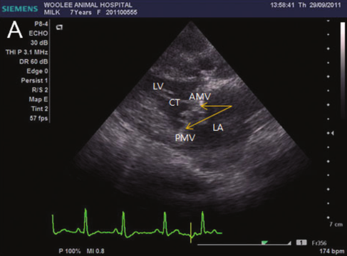


2D and color Doppler echocardiography in dogs with CMVI. (A) Mitral valve prolapse, which is characterized by one or both leaflets bent back into the left atrial chamber during systole, occurs commonly in dogs with CMVI. The severity of mitral valve prolapse was significantly correlated with MR severity. (B) The mitral valve lesions associated with CMVI are large and irregular in advanced stage of CMVI. Anterior leaflet of mitral valve is more commonly affected than posterior leaflet in dogs. (C) Color‐flow Doppler imaging in 2D echocardiography revealed severe regurgitant jets from left ventricle to left atrium during systole and is widely used for detection and assessment of MR in dogs with CMVI. (D) The MR can be also detected in color M‐mode echocardiography on the LV short‐axis view. LV, left ventricle; CT, chordae tendineae; AMV, anterior mitral valve; PMV, posterior mitral valve; RV, right ventricle; RA, right atrium.
Sang-II Suh, Dong-Hyun Han, Seung-Gon Lee, Yong-Wei Hung, Ran Choi and Changbaig Hyun (December 21st 2016). Chronic Mitral Valve Insufficiency in Dogs: Recent Advances in Diagnosis and Treatment, Canine Medicine - Recent Topics and Advanced Research, Hussein Abdelhay Elsayed Kaoud, IntechOpen, DOI: 10.5772/65689.


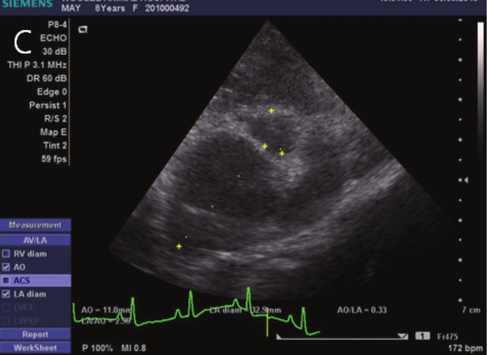

2D and M‐mode echocardiography in dogs with CMVI. (A) Abnormal excursion (decreased EF slope) and thickening of anterior mitral leaflet can be detected in M‐mode echocardiography. (B) The eccentric hypertrophy, which is characterized by an increase in end‐diastolic left ventricular dimensions (EDV), occurs in dogs with CMVI. (C)–(D) Hemodynamically significant chronic MR can induce volume overload, which subsequently can increase LV and LA volume and can result in LA and LV dilation. The degree of left atrial enlargement that is assessed by the left atrium to aorta (LA/Ao) ratio in 2D and M‐mode echocardiography and is closely correlated with the severity of heart failure.
Sang-II Suh, Dong-Hyun Han, Seung-Gon Lee, Yong-Wei Hung, Ran Choi and Changbaig Hyun (December 21st 2016). Chronic Mitral Valve Insufficiency in Dogs: Recent Advances in Diagnosis and Treatment, Canine Medicine - Recent Topics and Advanced Research, Hussein Abdelhay Elsayed Kaoud, IntechOpen, DOI: 10.5772/65689.


Pulse and continuous Doppler and tissue Doppler echocardiography in dogs with CMVI. (A) The transmitral flow profile consists of E and A and is affected by the pressure gradient between the LA and LV. Elevated E represents increased LA pressure and a worsening of heart failure. (B) Continuous Doppler echocardiography is useful to detect MR in dogs with CMVI. However, the degree of MR is not correlated with the severity of CMVI. (C) Pulse Doppler echocardiography in pulmonary venous flow is also useful to assess the progression of CMVI. The presence of pulmonary venous flow at atrial systole (PVa) indicates high LA pressure noticed in advanced stage of CMVI. (D) The early mitral inflow velocity to early mitral annular tissue velocity (E:Ea) ratio can be used to assess LV diastolic function. The E:E’ ratio is significantly correlated with left ventricular filling pressures.
Sang-II Suh, Dong-Hyun Han, Seung-Gon Lee, Yong-Wei Hung, Ran Choi and Changbaig Hyun (December 21st 2016). Chronic Mitral Valve Insufficiency in Dogs: Recent Advances in Diagnosis and Treatment, Canine Medicine - Recent Topics and Advanced Research, Hussein Abdelhay Elsayed Kaoud, IntechOpen, DOI: 10.5772/65689.


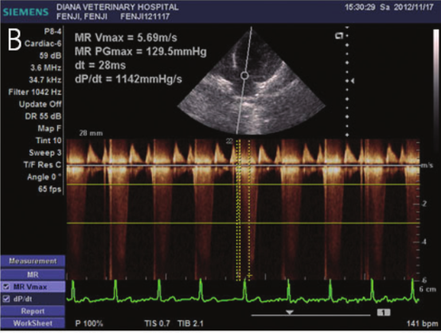
Determination of Doppler‐derived dP/dt and −dP/dt from the CW Doppler spectrum of the MR jet obtained from a dog with MR.
Kim JH, Park HM. "Usefulness of conventional and tissue Doppler echocardiography to predict congestive heart failure in dogs with myxomatous mitral valve disease". J Vet Intern Med. 2014;29(1):132–140. doi:10.1111/jvim.12466


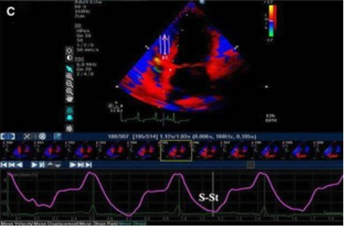
Radial tissue Doppler velocity (A), SR (B), and St (C) of the LV wall in a control dog. Note the ROI was positioned between the papillary muscles on the right parasternal short‐axis view. S, systolic velocity; E, E wave velocity; A, A wave velocity; SR, strain rate; St, strain; ROI, region of interest; LV, left ventricle.
Kim JH, Park HM. "Usefulness of conventional and tissue Doppler echocardiography to predict congestive heart failure in dogs with myxomatous mitral valve disease". J Vet Intern Med. 2014;29(1):132–140. doi:10.1111/jvim.12466


Longitudinal basal tissue Doppler velocities (A), SR (B), and St of the IVS wall in a control dog. Note the ROI was placed on the basal or apical region within the IVS and LV walls on the left parasternal apical 4‐chambered view. Arrows represent sample segments located within the LV and IVS walls. SR, strain rate; St, strain; IVS, interventricular septum; ROI, region of interest; LV, left ventricle.
Kim JH, Park HM. "Usefulness of conventional and tissue Doppler echocardiography to predict congestive heart failure in dogs with myxomatous mitral valve disease". J Vet Intern Med. 2014;29(1):132–140. doi:10.1111/jvim.12466
